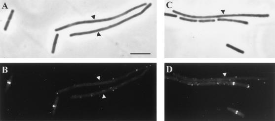
FIG. 2.
DivIB localization in SU391 (Pspac-RRC) and SU392 (Pspac-divIC) in the presence of IPTG at 45°C. All panels show both SU391 and SU392 cells (long and short, respectively) that were mixed prior to IFM processing. (A and C) Phase-contrast images; (B and D) fluorescein isothiocyanate-immunostained images of the same field. Cells were grown at 30°C to mid-exponential phase and then shifted to 45°C for 45 min. Arrowheads indicate the long SU391 cells. Bar = 5 μm (A).

