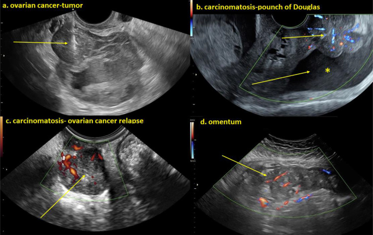
Figure 1.
(a) Heterogeneous irregular cystic-solid ovarian tumor and the tru-cut biopsy needle visualized at sampling of the solid component of the lesion. (b) Irregular solid richly vascularized carcinomatosis deposits on peritoneal surface and free fluid *In the pouch of Douglas (c) Heterogeneous solid richly vascularized metastatic tumor relapse in the pouch of Douglas. (d) Irregular solid heterogeneous moderately vascularized omentum metastasis of ovarian cancer.

