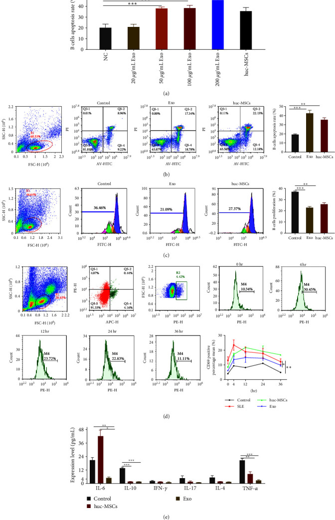
Figure 3.

hucMSCs-Exo promoted B cell apoptosis, inhibited proliferation, and prevented overactivation and inflammation. (a) Different concentrations of Exo were cocultured with B cells from SLE. (b) B cell apoptosis was determined using Annexin V-FITC kit (c) B cell proliferation was determined using the CFSE kit. (d) The expression of B cell activation marker CD69. (e) Cytokine expression levels after coculture with hucMSCs and exosomes compared with control. Exosomes were isolated from hucMSC. Data are expressed as the mean ± SEM. ∗P < 0.05, ∗∗P < 0.01, ∗∗∗P < 0.001.
