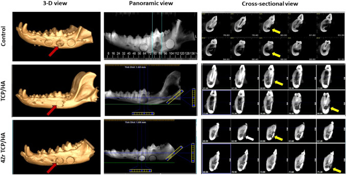
Fig. 2.
Three dimensional, panoramic and cross-sectional view cone-beam computed tomography of the induced mandibular defects at 12 weeks examination. Control defects (red arrow) demonstrated radiolucent circular defect with limited peripheral radiopacity without buccal rim corticalization (yellow arrow) around inferior alveolar canal (IAC). Defects grafted with tricalcium phosphate hydroxyapatite scaffolds (TCP/HA) (red arrow) demonstrated reduction in the size of the defect that was bounded by new bone formation with corticalization of the buccal rim (yellow arrow) around IAC. Defects grafted with zirconia-doped tricalcium phosphate hydroxyapatite scaffolds (4Zr TCP/HA) (red arrow) demonstrated reduction intimate contact with new bone formation with buccal rim corticalization around IAC (yellow arrow) with subperiosteal new bone formation (white arrow)

