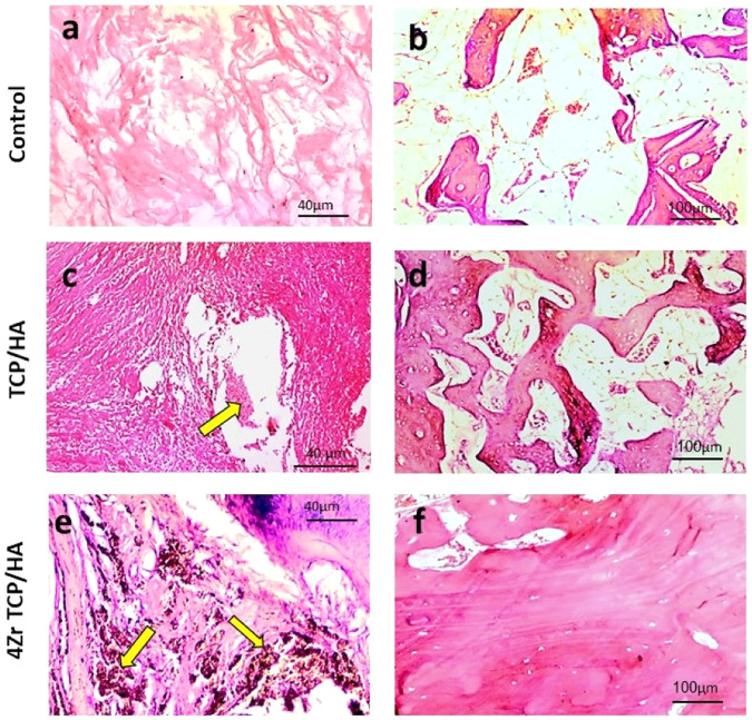
Fig. 3.
Photomicrograph of the mandibular defects in the studied groups stained with H&E. In the control group, the defect areas showed loose connective tissue (a) and osteoid tissue formation (b). In the TCP/HA group, the defects showed remnants of the graft material (yellow arrow) within a more condensed fibrous connective tissue (c), sections also showed newly formed haphazardly arranged bone trabeculae within a fatty marrow and blood vessels (d). In 4Zr TCP/HA group, remnants of the graft material (yellow arrows) were seen surrounded by dense fibrous connective tissue (e), also more organized bone tissue was evident with bone marrow, blood vessels and osteoblastic proliferation (f)

