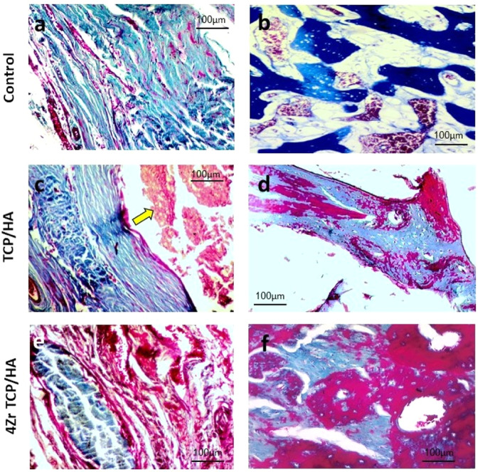
Fig. 4.
Photomicrograph of the mandibular defects in the studied groups stained with Masson trichrome. The control group showed loose connective tissue and immature bone (a) and formation of few spicules of osteoid bone tissue (b). The TCP/HA group revealed presence of remnants of the graft material (yellow arrow) in the defect site surrounded by connective tissue and newly formed bone (c), newly formed bony trabeculae showing slight maturation, with osteoblasts on their periphery and osteocytes in their lacunae (d). The 4Zr TCP/HA group showed the graft material enclosed within thin layer of fibrous connective tissue and blood vessels engorged with red blood cells (e), and showing noticeable bone maturation with more organized bone tissue, osteocytes in their lacunae and Haversian canals (f)

