
Fig. 2. ATUX-3364 or ATUX-8385 decreased hepatoblastoma proliferation.
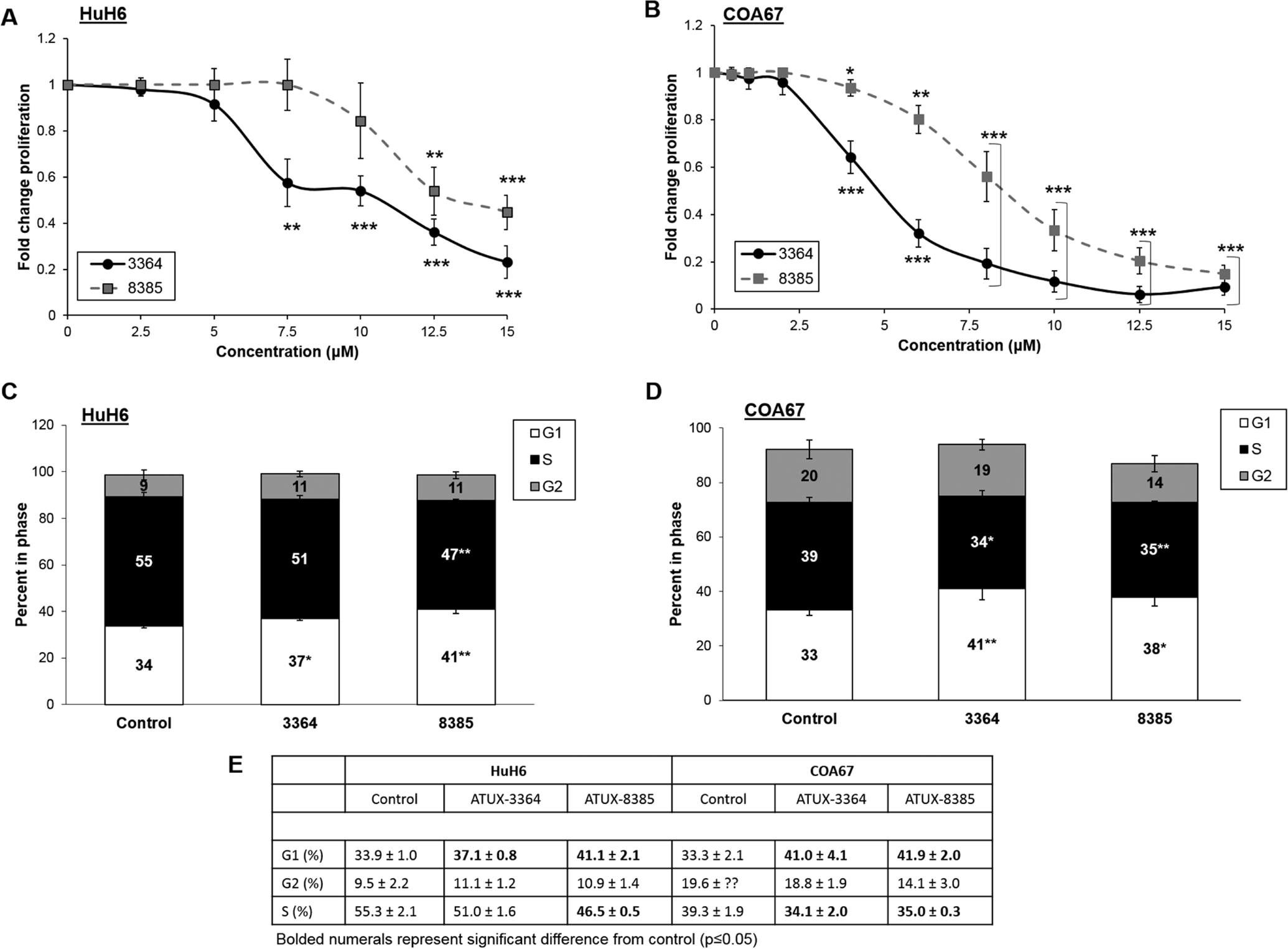
HuH6 or COA67 cells (5 × 10³ cells) were plated into 96-well plates and treated for 24 h with increasing doses of 3364 or 8385. (A) In HuH6 cells, 3364 and 8385 significantly decreased proliferation. (B) In COA67 cells proliferation was significantly decreased after treatment with either compound. (C) HuH6 cells were serum starved overnight and treated for 24 h with 3364 (6 μM) or 8385 (8 μM). There was a significant increase in the percentage of cells in G1 phase following treatment with either compound. There was an associated significant decrease in S phase following 8385 treatment with a trend toward significance for cells treated with 3364, indicating a lack of progression through the cell cycle. (D) Similarly, following 24 h treatment of 3364 or 8385 (4 μM), COA67 cells had a significant increase in percentage of cells in G1 phase and decrease in S phase, indicating a lack of progression through the cell cycle. (E) Cell cycle data presented in tabular form. Significant values (p≤0.05) depicted by bold font. Proliferation data reported as mean fold change ± SEM, cell cycle data reported as the mean percent cells in phase ± SEM. Data evaluated with two-tailed t-test. Experiments were repeated with at least three biologic replicates. *p≤0.05, **p≤0.01, ***p≤0.001.