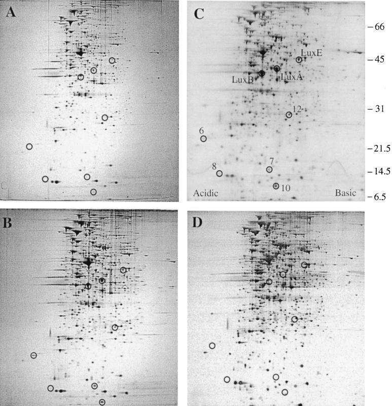
FIG. 1.
Production of QSR proteins in V. fischeri. Whole-cell proteins were analyzed by 2-D PAGE (Materials and Methods) for (A) MJ-100 (parent strain) at mid-exponential phase (A600 = 0.4), (B) MJ-100 at late exponential phase (A660 = 0.8), (C) MJ-100 at early stationary phase (A660 = 1.2), and (D) MJ-215 (ΔluxI ainS) at early stationary phase (A660 = 1.2). (C) The positions of LuxA, LuxB, LuxE, and the newly identified QSR proteins QSR 6 (AcfA), QSR 7 (unidentified), QSR 8 (QsrV), QSR 10 (QsrP), and QSR 12 (RibB) are circled and designated. The positions of molecular size standards are indicated at the right (in kilodaltons).

