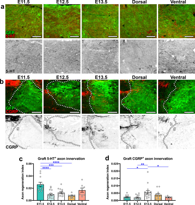
Fig. 5. Host serotonergic axons preferentially regenerate into earlier-stage grafts and host nociceptive axons preferentially regenerate into later-stage grafts.
Images of GFP+ grafts at 4 weeks post-transplantation, showing regeneration of a host 5-HT+ axons and b host CGRP+ axons into grafts. The bottom row of each panel shows axons in grayscale. b Host CGRP+ fibers are shown at the upper left of each image; each field of view contains the graft/host border (dotted lines) with point of entry of CGRP+ axons into grafts (g). c, d Quantification of c 5-HT+ axon density and d CGRP+ axon density within grafts. *P < 0.05, **P < 0.01, ***P < 0.001, ****P < 0.0001 by one-way ANOVA with Tukey’s multiple comparisons test. E11.5 (n = 11); E12.5 (n = 16); E13.5 (n = 16); dorsal (n = 12); ventral (n = 8). All data are mean ± SEM. Scale bars = 100 μm. Source data are provided as a Source Data file. The experiments in panels a and b were performed twice with similar results.

