
Fig. 1. Synthetic antigen sensors enable precise localization and measurement of IgG binding avidity on crowded membrane surfaces.
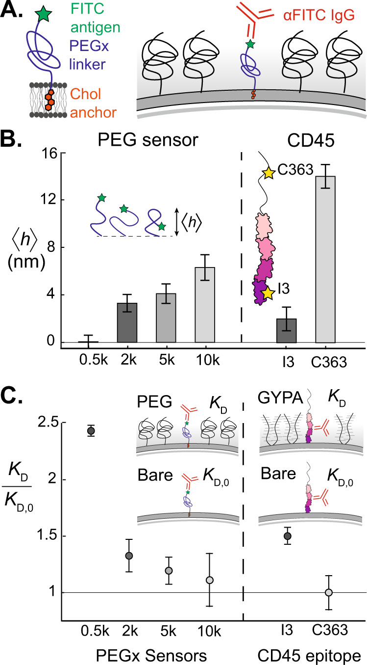
A Cholesterol-PEG-FITC sensors insert into the membrane and present the FITC antigen at varying heights above the membrane, depending on the PEG linker length. Sensors are exogenously inserted into reconstituted or live plasma membranes, and α FITC IgG avidity is a direct reporter of local crowding. B (Left) Mean height of synthetic antigen sensors increases as a function of PEG linker molecular weight, as measured by CSOP31. Error bars represent standard error of the mean for n = 65, 81, 79, and 54 beads, respectively. (Right) Epitope heights of two different α CD45 antibodies, α I3 and α C363 on the transmembrane tyrosine phosphatase CD45, as reported by Son et al.31. Error bars correspond to ~±1 nm error, reported by Son et al. C (Left) Dissociation constants of α FITC to the synthetic antigen sensors on a supported lipid bilayer, containing 3% DOPE-PEG2k as a surface crowder. Dissociation constants are normalized by the bare membrane value, KD,0. (Right) Two α CD45 antibodies with distinct epitope heights experience a significant difference in normalized avidity on a reconstituted membrane crowded with a mucin-like glycoprotein, Glycophorin A (GYPA). Points are derived from binding isotherm curve fits, and error bars are derived by fitting curves to data points 1 standard error of the mean above and below each data point, propagated with the 95% CI error of the fits. Curve fits for KD,0 are generated from populations of n = 912, 905, 1062, 635, 1730, and 1787 beads, respectively. Curve fits for KD are generated from populations of n = 2115, 1281, 1237, 578, 2180, and 3333 beads, respectively.