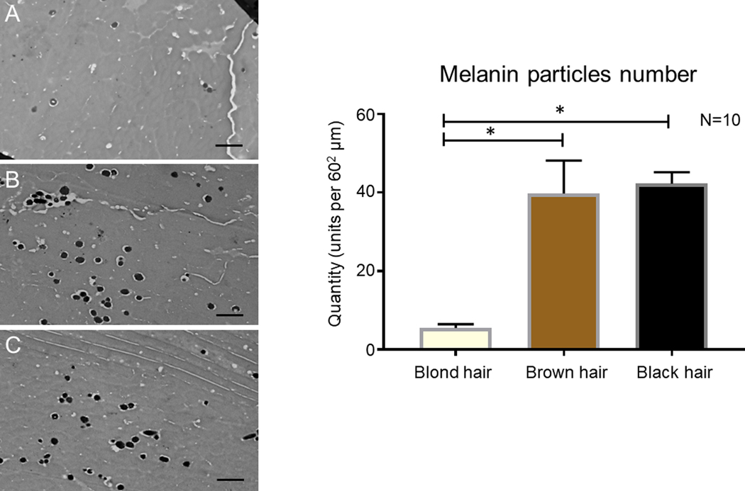
Figure 1.
Left panel: TEM Imaging of Melanosomes in Hair Shaft Slices: representative images of middle layer of hair shaft slices of: A–blond hair, B–brown hair, C–black hair. Right panel: Graph representing the quantity of melanosomes on the standard 2900X TEM slices from samples of blond, brown and black hair. Scale bar for TEM: 1 µm.

