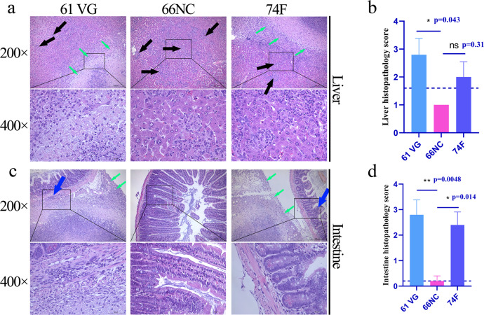
Fig. 6. Histopathological findings of the liver and intestine.
Histopathology was evaluated in the liver and intestine sections stained with hematoxylin and eosin from mice challenged by intraperitoneal injection with MAP K-10 for 2 weeks after vaccination. An image represents one experiment with at least five independent replicates. a Histopathological changes in the liver (scale bars, 200 μm (top) and 50 μm (bottom)). Montanide ISA 61 VG (61 VG): macro-granulomas (green arrow) and multiple microgranulomas (black arrow), 66NC: multiple microgranulomas (black arrow), 74 F: macro-granulomas (green arrow) and multiple microgranulomas (black arrow). The bottom images are enlarged from the outlined areas of the top images. b Liver histopathology score. c Histopathological changes in the intestine (scale bars, 200 μm (top) and 50 μm (bottom)). Montanide ISA 61 VG (61 VG): macro-granulomas (green arrow) and inflammatory cell infiltration (blue arrow), 66NC: without pathologies, 74 F: macro-granulomas (green arrow) and inflammatory cell infiltration (blue arrow). The bottom images are enlarged from the outlined areas of the top images. d Intestine histopathology score. b, d Mean ± SEM and one-way ANOVA were used to analyze statistical significance followed by Tukey’s multiple comparisons tests. ns, non-significant; *P < 0.05; **P < 0.01.

