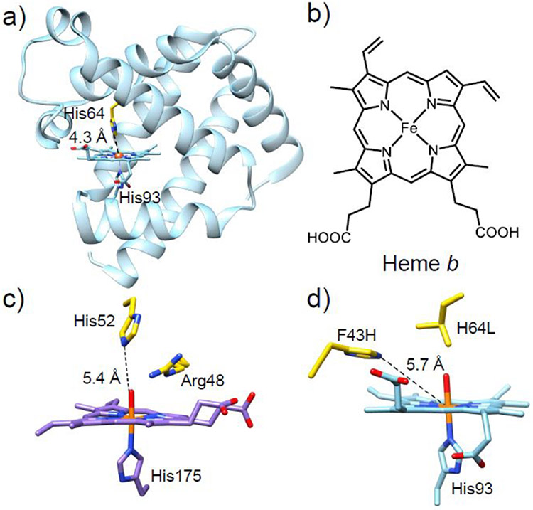
Figure 1.
(a) Crystal structure of WT-sperm whale Mb (swMb) (PDB ID: 5YCE).89 (b) Chemical structure of heme b. (c) Crystal structure of the active site of cytochrome c peroxidase (PDB ID: 1ZBY) with the distal His52 and Arg48 residues shown in yellow.90 (d) Crystal structure of the active site of the F43H/H64L Mb mutant (PDB ID: 1OFK)102 with the two mutated residues shown in yellow. The distances between the Nε atom of the distal His residue and the heme iron in each crystal structure are labeled.

