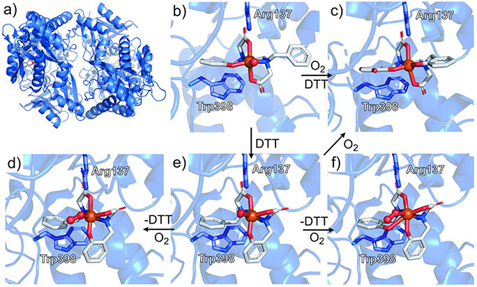
Figure 22.
(a) Crystal structure of NikA with bound FeIIIL0. (b) FeIIIL0 binding site of NikA with interacting SCS residues Arg137 and Trp398 highlighted in blue. (c) Dihydroxylated product formed following exposure of NikA/FeL0 to 50 mM DTT and O2. (d) O2-bound intermediate formed following four successive back soaks of DTT-reduced NikA/FeL0 in DTT-free mother liquor, followed by O2 exposure. (e) NikA/FeL0 soaked in 50 mM DTT. (f) Partially hydroxylated O2-bound intermediate formed following three successive back soaks of DTT-reduced NikA/FeL0 in DTT-free mother liquor, followed by O2 exposure. Respective PDB IDs for parts a—f: 3MVW, 3MVW, 3MW0, 3MVY, 3MVX, 3MVZ.302 Atom coloring: N (blue); O (red); Fe (orange).

