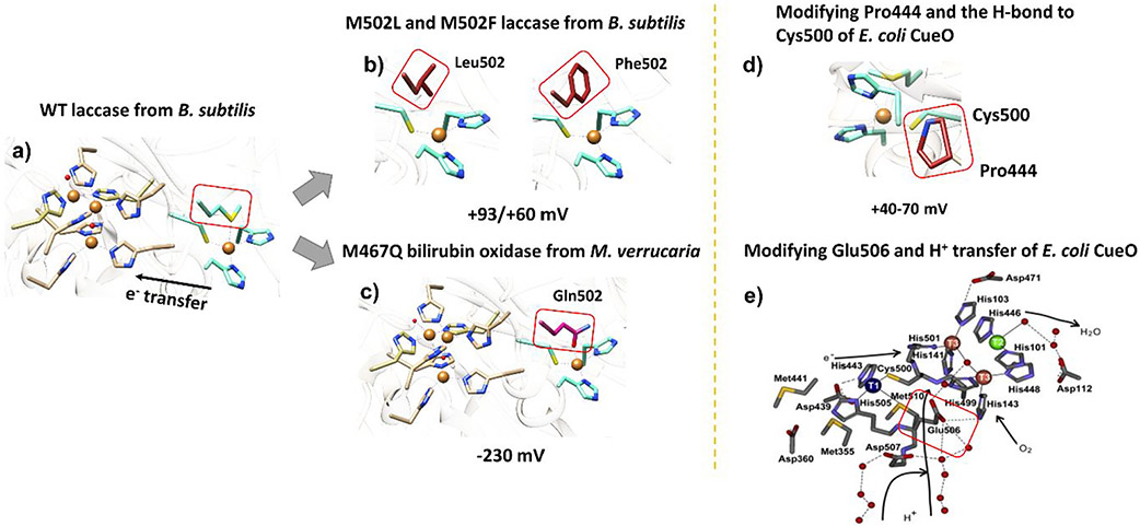
Figure 31.
(a) Structure of wild-type laccase from B. subtilis (PDB ID: 1GSK). The black arrow shows the electron transfer from the T1Cu to the T2/T3Cu site to reduce O2. (b) Axial mutants of B. subtilis laccase modeled from the wild-type (PDB ID: 1GSK)405 using UCSF Chimera.406 (c) M467Q axial mutant of bilirubin oxidase from M. verrucaria (PDB ID: 6IQX).407 (d) Modifying Pro444, the H-bond donor to the Cys residue of T1Cu in E. coli CueO (PDB ID: 1KV7).408 (e) Modifying Glu506, a proton transfer mediator residue in E. coli CueO. Reprinted with permission from ref 409. Copyright 2012 Elsevier.

