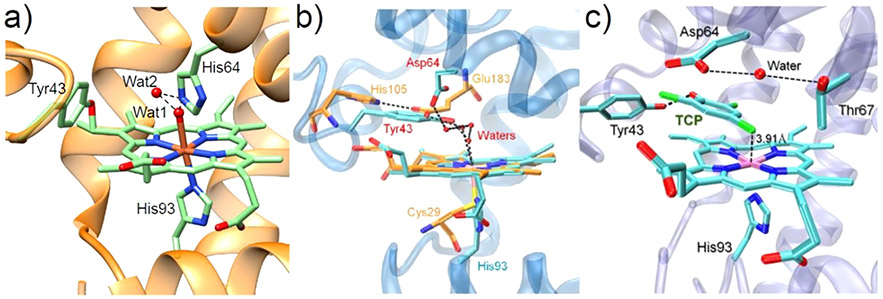
Figure 6.
(a) Crystal structure of F43Y Mb (PDB ID: 4QAU)115 showing the Tyr-heme covalent C─O bond and two distal water molecules forming H-bonding interactions (dotted lines).115 (b) Overlay of the crystal structures of ferric F43Y/H64D Mb (blue, PDB ID: 5ZZF)118 and the heme active site of native chloroperoxidase (orange, PDB ID: 1CPO),119 showing the H-bonding network. (c) Crystal structure of ferric F43Y/H64D Mb in complex with TCP (PDB ID: 5ZZG),118 showing the conformation of Tyr43 and TCP and the H-bonding interactions in the heme center. The distance between the Cl4 atom and the heme Fe (3.91 Å) is indicated. Reproduced with permission from ref 118. Copyright 2018 American Chemical Society.

