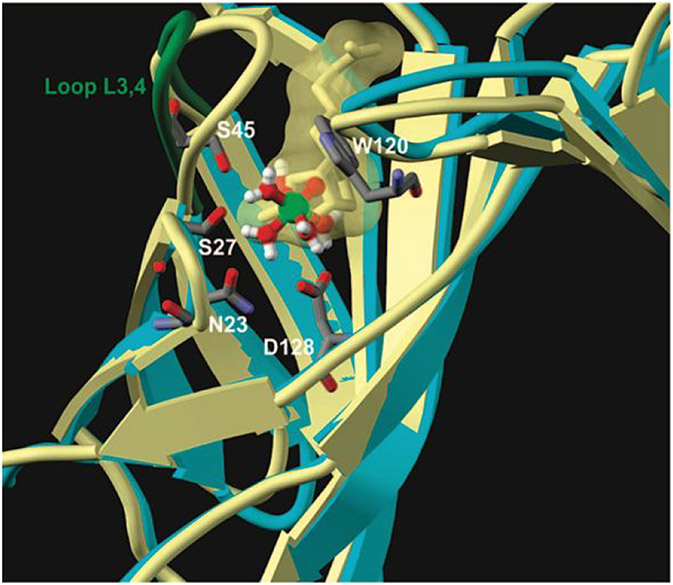
Figure 68.
Superimposition of the docked structure [VO(H2O)5]2+ (ball-and-stick representation) with WT-Sav (monomers A and D, light blue schematic secondary structure) with the structure of biotin with WT-Sav (PDB ID: 2IZG): biotin (yellow stick and yellow transparent surface); monomers A and D (yellow schematic secondary structure). Reprinted with permission from ref 589. Copyright 2008 American Chemistry Society.

