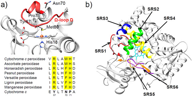
Figure 9.
(a) Crystal structure of yeast iso-1 Cc (top, PDB ID: 2YCC).147 The flexible coordination loop, Ω-loop D, is colored in red. At the bottom is the multiple sequence alignment of the distal heme region of some classical peroxidases which is compared to the sequence of the distal heme region of yeast iso-1 Cc.154 (b) Substrate recognition sites (SRSs) in P450 enzyme (CYP2C9, PDB ID: 1R9O)155 shown by arrows: SRS1 (red), SRS2 (green), SRS3 (blue), SRS4 (yellow), SRS5 (orange), SRS6 (magenta).156

