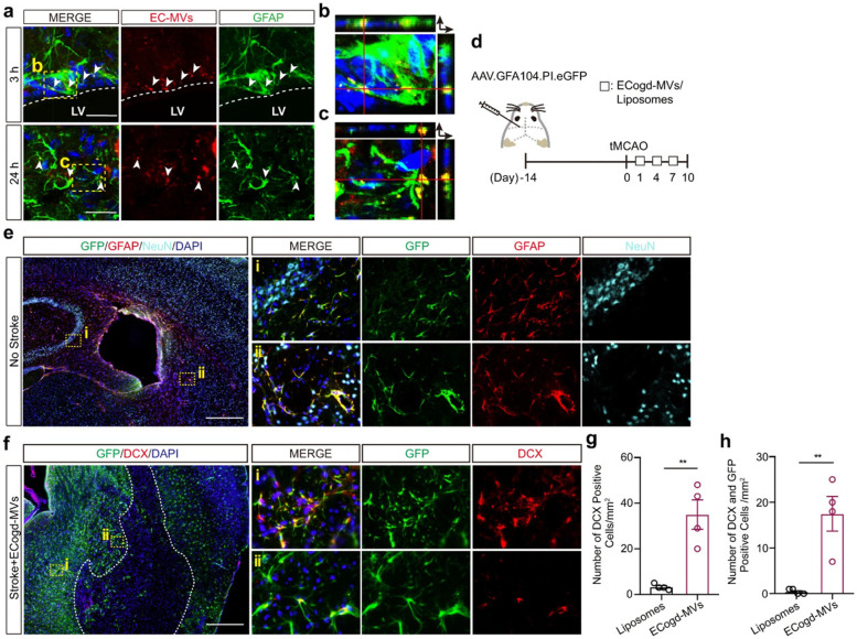
Fig. 6.
Hypoxia and hypoglycemia-treated ECs produce microvesicles (EC-MVs) with the potential to induce astrocyte differentiation toward neural progenitor cells in a mouse model of ischemia (a-h). Confocal images revealed the successful entry of red color Cy3-labeled EC microvesicles (EC-MVs) by astrocytes after 3 (a, b; Scale bar: 25 μm) and 24 h (a, c; Scale bar: 25 μm) after injection into the lateral ventricle (White color dash lines: lateral ventricle border). Schematic of experimental procedure (d). The expression of GFP, GFAP, and NeuN 14 days after administration of AAV.GFA104.PI.eGFP vector (e; Scale bar: 200 μm). In panel f, GFP and DCX positive cells were monitored on day 10 with transient MCAO (Scale bar: 200 μm). The number of DCX+ neurons/mm2 was measured in the peri-infarct zone (g) ROIs were calculated per each Sect. (3 sections per animal; each in 4 mice; p = 0.0026). DCX+/GFP+ cells per mm2 of in peri-infarct zone (3 sections per animal; each in 4 mice; p = 0.0043) (h). Three (a) and four (e, f) sets of experiments were performed in this study. Abbreviation: Doublecortin: DCX; Glial fibrillary acidic protein: GFAP; Green fluorescent protein: GFP; Neuronal nuclei: NeuN; Regions of Interests; ROIs. Reprinted with permission, [94] Copyright 2022. Nature Communications

