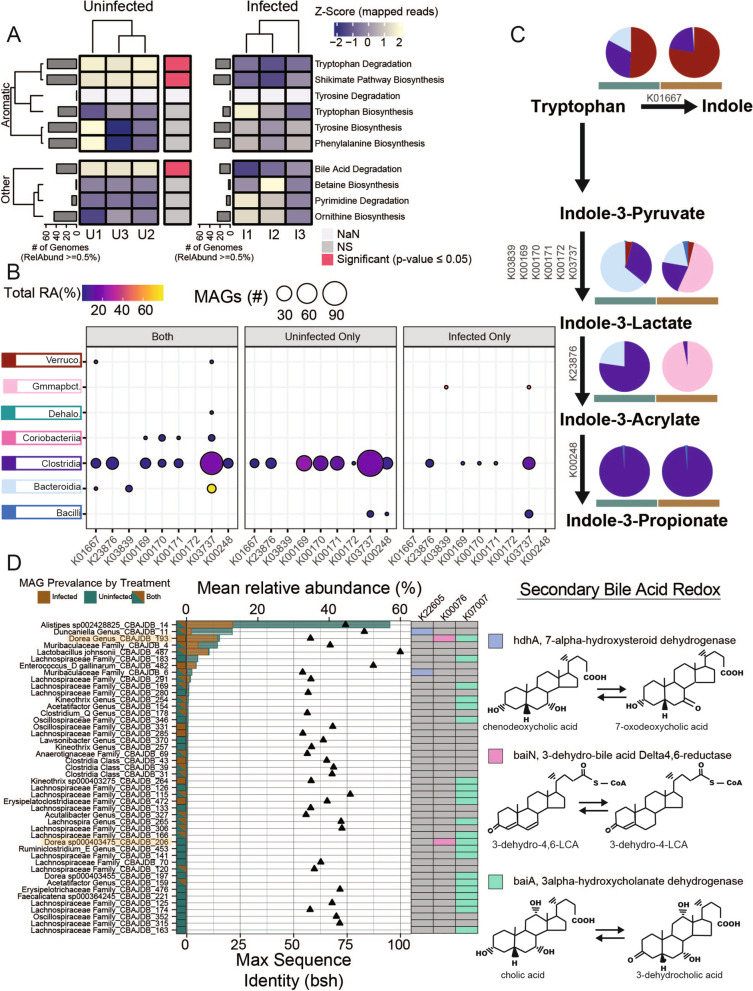
Fig. 6.
Tryptophan and bile acid metabolism in inflamed and uninfected gut microbiomes. A Mean relative abundance summed for each function (rows). Functions that are significantly different between treatments as determined by analysis of variance (ANOVA) (p ≤ 0.05) are indicated by horizontal bar between heatmaps with red highlighting significance at the function level. Gray bars indicate the number medium- and high-quality (MQHQ) metagenome assembled genomes (MAGs) that comprise at least 0.5% of the community with a given function. B Relative abundance (point color) and number of MAGs (size) in each class with each gene for tryptophan degradation separated by MAG presence in each treatment, where both indicate MAGs that recruited strictly mapped reads from both treatments. C Tryptophan degradation to indole and indole derivatives pathway with pie charts colored by proportion of MAGs in each class (coloring from B) for each treatment. D Relative abundance (bars) of MAGs in each treatment encoding bile salt hydrolase (bsh), points show sequence similarity to bsh, or hdhA (K22605), baiN (K00076), or baiA (K07007) involved in secondary bile acid metabolism. Dorea are highlighted as MAGs with more than one gene for metabolizing secondary bile acid products

