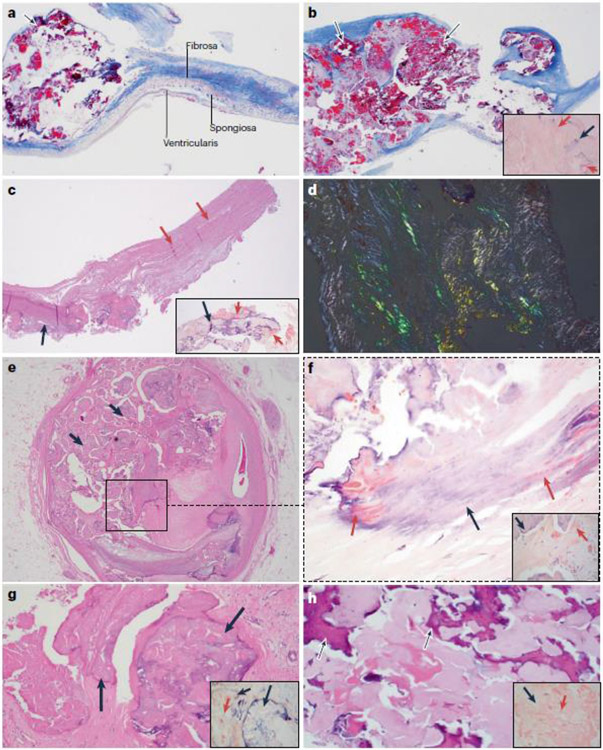
Fig. 1. Association between amyloid deposits and calcification.
a–d ∣ Calcification in native and bioprosthetic aortic valves is associated with amyloid deposits. Parts a and b are from a man aged 58 years with a bicuspid aortic valve who underwent aortic valve replacement for symptomatic aortic stenosis. The patient never smoked and he had no history of coronary artery disease, obesity, chronic kidney disease, diabetes mellitus or hypertension; a plasma lipid profile was not available. Part a shows trichrome staining of the excised aortic valve, revealing small deposits of nodular calcification (arrow) in the cusp (2× magnification). Adjacent is the layered architecture of the valve. On the aortic side is the fibrosa, which is rich in collagen fibres and provides tensile strength to the valve, and on the ventricular side is the ventricularis, which is rich in elastic fibres. Between the two layers is the spongiosa, which is primarily composed of proteoglycans. The valvular endothelial cells line both sides of the cusp. As seen in this image, the calcification starts in the fibrosa. Part b shows an area of calcification in the same valve, which now involves the full thickness of the cusp (arrows). In the inset image, Congo red staining for amyloid shows salmon-pink deposits of amyloid (red arrows) in the vicinity of calcification (black arrow). Part c is from a man aged 56 years who underwent aortic valve replacement with a bovine bioprosthetic valve 15 years ago and who recently presented to the hospital for evaluation of gradually worsening exertional dyspnoea over the past 2 years. The patient had no coronary risk factors and no history of major adverse cardiac events. A transthoracic echocardiogram showed severe aortic stenosis of the bioprosthetic valve, and he underwent aortic valve replacement again. Haematoxylin and eosin (H&E) staining of the excised aortic valve shows areas of nodular calcification (black arrow) that involves the full thickness of the acellular bioprosthetic valve (pink arrows) (4× magnification). In the inset image, Congo red staining shows salmon-pink deposits of amyloid (red arrows) adjacent to the calcified area (black arrow). Part d shows the apple-green birefringence that is characteristic of amyloid when examined by polarization microscopy using a polarizer and an analyser in the bioprosthetic valve (Congo red staining; 10× magnification). e–h ∣ Calcification at sites other than the aortic valve is also associated with amyloid deposits. Parts e and f are from a patient who underwent heart transplantation for severe coronary artery disease. Part e shows H&E staining of a section of the left main coronary artery that has fibrocalcific plaque with nodular calcification (black arrows) (2× magnification). Part f shows Congo red staining of the region in the square in part e, revealing salmon-pink amyloid deposits (red arrows) within the calcified areas (black arrow) and in the vicinity of calcification (inset image) (10× magnification). As the calcification increases, the salmon-pink deposits of amyloid seem to decrease. Part g shows H&E staining of a mitral valve from a man aged 79 years with mitral regurgitation due to mitral annular calcification and mitral valve prolapse. Nodular areas of calcification (black arrows) are present (10× magnification). In the inset image, Congo red staining shows salmon-pink deposits of amyloid (red arrow) within and adjacent to the calcified area (black arrows). Part h is from a woman aged 70 years with multiple calcified nodules in the lung, as seen on a CT scan. H&E staining of a biopsy sample from one of the nodules is positive for amyloid with areas of calcification (black arrows) (10× magnification). In the inset image, Congo red staining highlights salmon-pink deposits of amyloid (red arrow) and adjacent calcification (black arrow). In all the patients, the classic apple-green birefringence was seen with polarizing microscopy. Liquid chromatography and mass spectrometry (LC-MS) to subtype the amyloid deposits showed that the amyloid associated with the calcified valves (a-d and g) contained the signature proteins apolipoprotein A-IV, apolipoprotein E and serum amyloid P. Whereas the patients with valvular amyloid did not have one specific amyloid protein, LC-MS revealed that the amyloid found in the lung nodule (part h) was amyloid immunoglobulin λ-light chain. LC-MS was not performed on the amyloidosis present in the coronary artery calcification (parts e,f).

