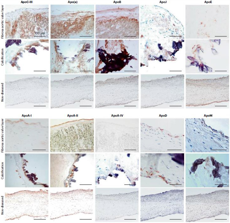
Fig. 2. Apolipoproteins in human aortic valve leaflets.
Aortic valve leaflets obtained from valve replacement surgeries were used for mass spectrometric analysis. Three areas were dissected from eachs leaflet: the calcific and fibrotic areas, and the area with no discernible pathology (referred to as non-diseased). The tissues were lysed and the protein constituents proteolyzed for first unbiased label-free mass spectrometry (modified from reference 32), and then for labeled-based targeted mass spectrometry (modified from reference 28), both performed on a benchtop quadrupole Orbitrap mass spectrometer (Q Exactive). The initial unbiased proteome profiling demonstrated that several apolipoproteins were present in aortic valve leaflets, most of which were enriched in the calcific portions: ApoA-I, ApoA-II, ApoA-IV, ApoB, ApoC-III, ApoD, ApoL-I and ApoM, all of which were verified using targeted mass spectrometry (Schlotter F, JBC 2021). While the other apolipoproteins, Apo(a), ApoE, ApoH and ApoJ were not differentially enriched in any leaflet area, they were nonetheless detected in all aortic valve leaflets.
This figure demonstrates representative immunohistochemistry images of aortic valve leaflets’ cryosections stained with antibodies against ApoCIII, Apo(a), ApoB, ApoJ, ApoE, ApoA-I, ApoA-II, ApoA-IV, ApoD and ApoM. The images show positive diffuse staining in the fibrosa layer (top panel, fibrosa facing up) for ApoCIII, Apo(a) and ApoB antibodies (red-brown reaction product) and cellular expression for ApoE, ApoA-I, ApoA-II, ApoA-IV, ApoD and ApoM. Most apolipoproteins are highly expressed around calcific nodules (middle panel). Non-diseased leaflets (bottom panel) show negative antibody reaction, except a weak staining for ApoE, ApoA-I and ApoA-II. Our mass spectrometric and immunohistochemistry analyses indicate that valves from patient with calcific aortic stenosis are highly enriched in apolipoproteins, particularly localized to the calcification-prone fibrosa and calcific nodules, thus suggesting their contribution to calcification process. (Modified from reference 28).

