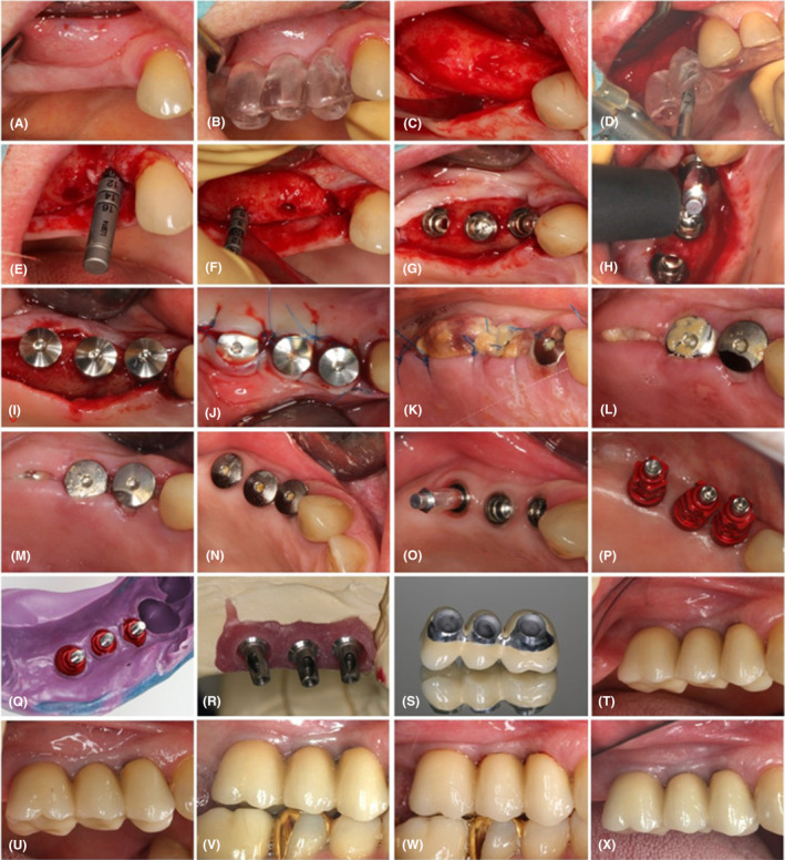
FIGURE 2.

Workflow of prosthetic rehabilitation. (A) Maxillary arch at baseline. (B) Surgical guide for dental implant placement. (C) Reflection of full‐thickness flap at the site if implant insertion. (D) Preparation of the implant beds using a surgical guide. (E) Validation of implant bed adequacy at edentulous site of former tooth no. 14. (F) Validation of implant bed adequacy at edentulous site of former tooth no. 16. (G) Insertion of implants at edentulous sites of former teeth no. 14, 15, and 16. (H) Measurement of implant stability immediately after insertion using Ostell® equipment (Integration Diagnostics). (I) Insertion of closure screws onto implants. (J) Suturing of the full‐thickness flaps. (K) Surgical site 4 days after implant insertion; secondary herpetic lesions on the palate. (L) Surgical site following suture removal, 14 days after implant insertion. (M) Surgical site 1 month after implant insertion. (N) Surgical site 6 months after implant insertion. (O) Measurement of implant stability 6 months after implant insertion using Ostell® equipment (Integration Diagnostics). (P) Positioning of open‐tray copings for the registration procedure. (Q) Implant position registration utilizing open‐tray copings and polyether impression material (Impregum®, 3 M ESPE). (R) Transfer of implant position onto a plaster cast. (S) The metal framework made of Coron®, a cobalt‐chromium alloy (Institut Straumann AG). (T) Prosthetic restoration after cementation. (U) Prosthetic restoration 1 year after cementation. (V) Prosthetic restoration 2 years after cementation. (W) Prosthetic restoration 3 years after cementation. (X) Prosthetic restoration 4 years after cementation.
