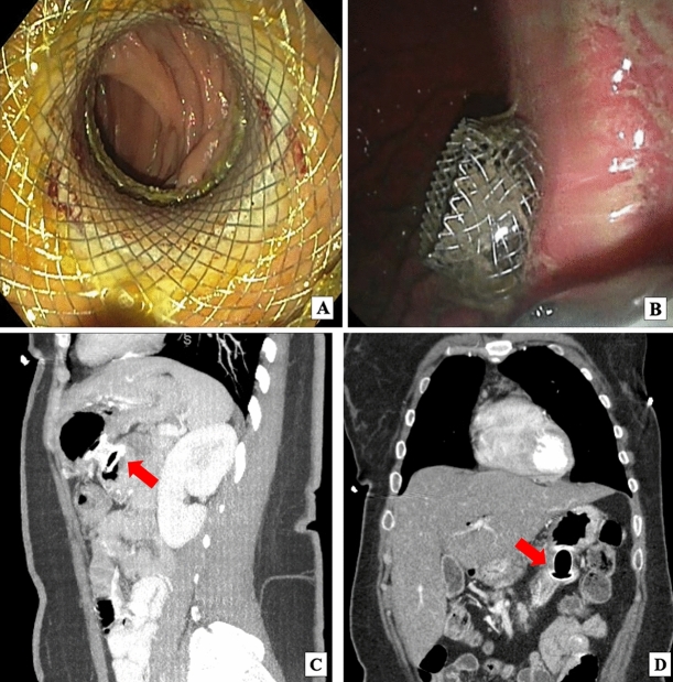
Fig. 4.
Endoscopic images of lumen apposing metal stent (LAMS) deployment between stomach and jejunum and abdominal CT images with intravenous contrast demonstrating LAMS gastrojejunostomy (GJ). A Jejunum as seen through the LAMS. After contrast was injected to identify the jejunal lumen distal to the duodenal stent that had become stenosed, a 20 mm × 10 mm electrocautery-enhanced LAMS (Hot Axios, BSCI) was deployed from the stomach to the small bowel just distal to the duodenal stent. The LAMS was dilated to 18 mm using a wire-guided balloon dilator. Small bowel was examined distal to the LAMS which was healthy. No trauma from the LAMS was demonstrated. B Deployment of LAMS flange in the stomach. The distance between the gastric and jejunal wall was less than 1 cm. There were no significant blood vessels in the path chosen for LAMS entry. C Sagittal abdominal CT with red arrow pointing to interval LAMS placed between posterior gastric wall and proximal jejunum, distal to the end of the obstructed duodenal stent. D Coronal abdominal CT with red arrow pointing to LAMS between stomach and proximal jejunum

