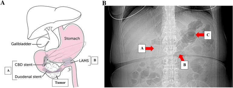
Fig. 5.

A Location of common bile duct (CBD), duodenal, and GJ stents relative to duodenal mass. A Cannulation of the bile duct, biliary sphincterotomy, and 8 mm × 60 mm fully covered self-expanding metal stent (FCSEMS, Viabil Fore) placement into biliary duct with distal end in duodenum were performed via ERCP. Circumferential, fungating, obstructing mass in the distal descending duodenum involving the major papilla causing 3 cm stricture was traversed with pediatric colonoscope under endoscopic and fluoroscopic guidance. A 22 mm × 90 mm uncovered self-expanding metal stent (USEMS, WallFlex BSCI) was placed across the duodenal stricture, with the proximal end at the level of the major papilla near the previously placed biliary stent. B LAMS was placed two and a half months later. B Abdominal X-ray with red arrows pointing to CBD (A), duodenal (B), and LAM (C) stents
