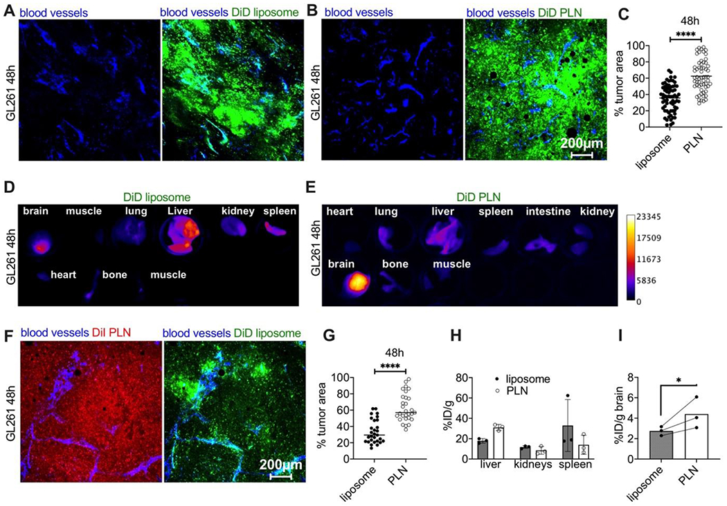
Fig. 3. PLN-formulated ICLs accumulate in gliomas at 48h more efficiently than liposomal ICLs.

A-B) Representative ex vivo confocal microscopy images (from 3 mice per group) of tumor slices from mice injected with liposomal DiD or PLN-formulated DiD. Scale bar is the same for all images; C) quantification of % fluorescent area in tumors (n=3 mice per group, pairwise t-test); D-E) pseudocolored images of DiD fluorescence in main organs after injection of liposomal DiD or PLN-formulated DiD; F) representative confocal microscopy images of tumor (central area) after co-injection of PLN-formulated DiI (Supplemental Fig. 2) and liposomal DiD; G) quantification of tumor’s fluorescent area after co-injection of DiI PLN and DiD liposomes (n=3 mice per group, pairwise t-test); H-I) accumulation (% ID/g) of liposomal and PLN-formulated ICLs after extraction from clearance organs and brains of GL261 tumor-bearing mice. N=3 mice, pairwise t-test. P-value: ****<0.0001; *=0.04.
