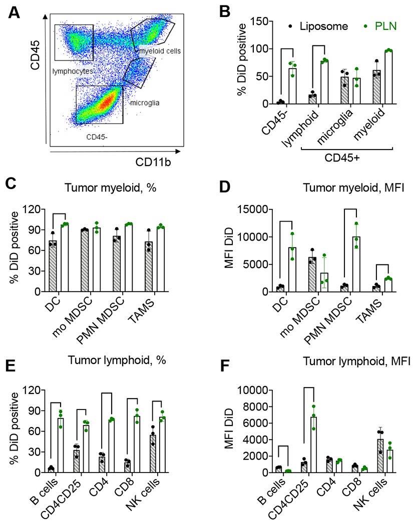
Fig. 5. Flow cytometry analysis demonstrates better accumulation of PLN-formulated DiD than liposomal DiD.

GL261 tumor-bearing mice were injected with DiD liposomes or DiD PLNs, and the cell uptake was analyzed 48h post-injection. A) Gating strategy (also Supplemental Figs. 4–6); B) CD45+ and CD45− populations (% or cells). Bar labels are the same for all subsequent graphs; C) myeloid cell subtypes (% or cells); D) myeloid cell subtypes (mean fluorescence intensity, MFI); E) lymphoid cell subtypes (% of cells); F) lymphoid cell subtypes (MFI). Dendritic cells (DC), monocytic MDSCs (mo MDSC), polymorphonuclear MDSC (PNM MDSC), and tumor-associated macrophages (TAMs). N=3 mice per group, parametric t-tests with multiple comparisons. Only significant comparisons are indicated. P-value: ****<0.0001; ***<0.001, **<0.01, *<0.05.
