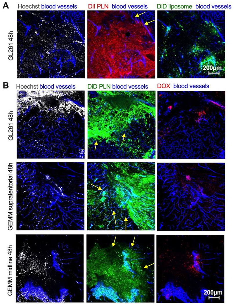
Fig. 6. Localization of PLN-formulated ICLs in tumor margin 48h post-injection.

Mice were injected and imaged as described in Fig. 3. A) DiI PLN and DiD liposomes were coinjected in GL261 mice. Difference in the margin (arrows) localization between PLN-formulated DiI and liposomal DiD in GL261 tumor; B) PLN-formulated DiD and PEGylated liposomal doxorubicin (DOX) were coinjected in GL261 mice (top row) and in genetically engineered invasive mouse models (supratentorial high-grade and diffuse midline glioma, middle and bottom rows). Arrows point to the margin. The size bar is the same for all images. The experiment is repeated twice.
