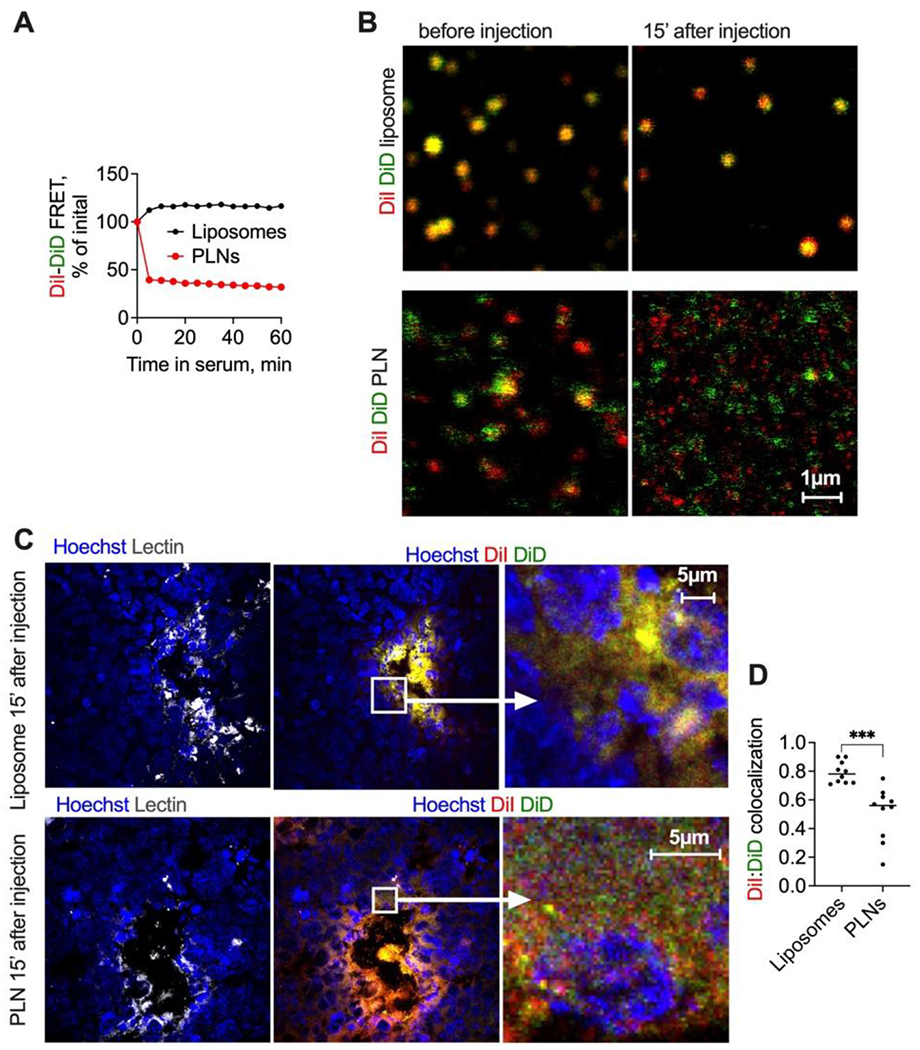
Fig. 8. PLNs show instability in serum in vitro and in vivo.

A) Normalized FRET efficiency (Supplemental Fig. 9 for absolute FRET values) after incubation of double-labeled PLNs and liposomes in mouse serum; B) confocal microscopy in plasma post-injection shows the disintegration of PLNs and stability of liposomes. The size bar is the same for all images; C) high magnification microscopy of GL261 tumor histological sections show a separation of PLN-formulated DiI/DiD, but not liposomal DiI/DiD at early steps of tumor entry; D) colocalization of DiI and DiD in PLN injected tumors and in liposome-injected tumors (Pearson coefficient, calculated with Coloc2 in Fiji).
