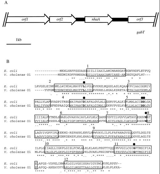
FIG. 1.
(A) Genetic organization of the nhaA locus of Vibrio cholerae O1. (B) CLUSTAL W alignment of E. coli and V. cholerae O1 NhaA proteins. Asterisks indicate amino acid identity, and dots indicate amino acid similarity. The conserved amino acid residues Asp-133, Asp-163, Asp-164, His-225, Leu-73, and G-338 are indicated with boldface circles. Helical structures spanning the membrane are indicated with open boxes and are numbered. The E. coli sequence data were taken from Taglicht et al. (48).

