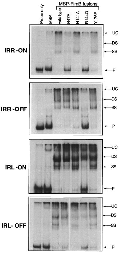
FIG. 5.
Electrophoretic mobility shift analysis of FimB-containing lysate interactions with IRR or IRL sequences from fimS. Data are shown for the IRR and IRL amplified from phase-on or phase-off fimS elements. Lanes 1, no added protein; lanes 2, MBP alone; lanes 3, MBP-FimB; lanes 4, MBP-FimB R47A; lanes 5, MBP-FimB H141A; lanes 6, MBP-FimB R144Q; lanes 7, MBP-FimB Y176F. Species corresponding to complexes with two bound protein protomers (DS) and a single bound protomer (SS) are shown. Also indicated are an unknown complex (UC) that migrated near the top of all lanes and the unbound radiolabeled probe (P). The IRL DNA fragment was 111 bp, while the IRR DNA fragment was 205 bp.

