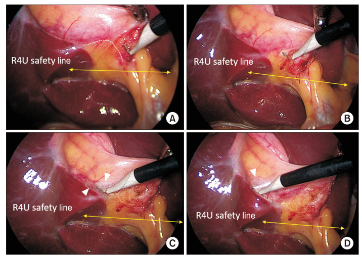
Fig. 3.
Dissection of the hepatocystic triangle from the lateral/right side (posterior dissection): Peritoneal fold division. (A) Peritoneal fold is divided close to the gallbladder (broken line). (B) Hook cautery tip is introduced beneath the peritoneal fold and gently elevated with side to side sweeping movements to open up the space and (C) to allow CO2 gas to enter beneath the peritoneal layer for facilitating (pneumo-) dissection (arrowheads), and (D) the peritoneal fold is then gradually divided towards the fundus. Dissection starts above the R4U safely line. To facilitate proper dissection, gallbladder infundibulum is retracted in left-cephalad direction.

