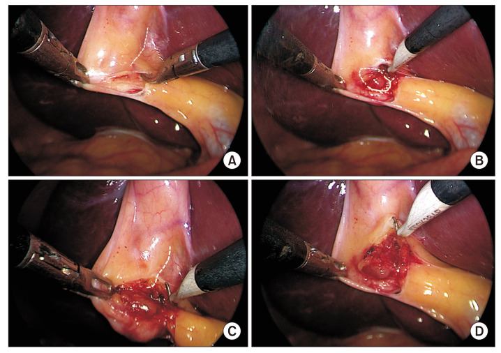
Fig. 4.
Dissection of the hepatocystic triangle from the medial/left side (anterior dissection): peritoneal fold division. After opening the peritoneal fold (see Fig. 2), (A) anterior peritoneal fold is gently dissected off the underlying tissue by blunt dissection followed by (B–D) its division close to the gallbladder (broken line) while exposing the cystic lymph node (marked with circle; broken line in ‘B’). To facilitate proper dissection, gallbladder infundibulum is retracted in right-caudal direction.

