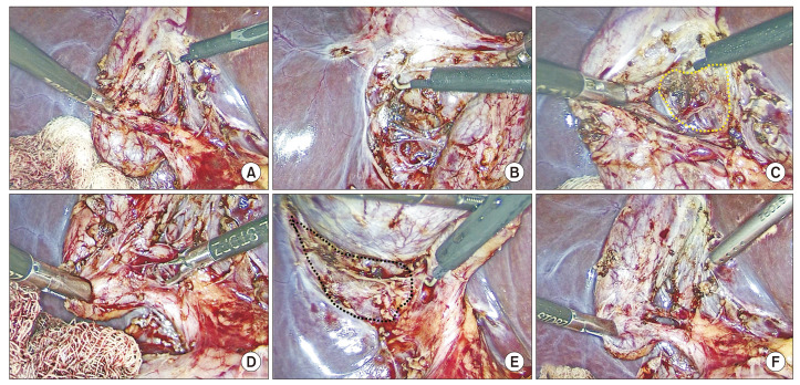
Fig. 6.
Dissection of the hepatocystic triangle and exposure of the cystic plate. Cystic plate is being exposed by the alternate dissection from (A) medial/anterior and (B) lateral/posterior aspects with (C, E) subsequent exposure of the necessary extent of the plate (marked with broken line). Calots’ triangle is subsequently dissected from (D) anterior and (E) posterior aspects to (F) finally achieve the critical view of safety (CVS). This figure also illustrates the cystic plate first approach to achieve the CVS (also see Supplementary Video 1).

