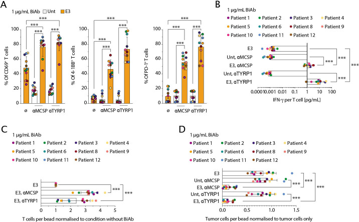
Figure 3.
αMCSP/αE3 and αTYRP1/αE3 BiAb activate SAR T cells to mediate specific cytotoxicity against patient-derived melanoma samples. (A) Human T cells were co-cultured with patient-derived melanoma samples (effector to target ratio 2:1) and either αMCSP/αE3 or αTYRP1/αE3 BiAb (αMCSP or αTYRP1, 1 µg/mL) for 48 hours. The frequency of CD69, PD-1 and 4-1BB on T cells was assessed using flow cytometry. (B) Supernatant was taken and analyzed with ELISA for IFN-γ.The values were normalized to the numbers of plated T cells. (C) The CD3+ T cell count per bead was measured and normalized to conditions without BiAb. (D) The percentage lysis of the patient-derived melanoma samples by SAR T cells and either of the two BiAb was calculated based on flow cytometric readout after 48 hours of co-culture. The values shown were normalized to the tumor cells only control conditions. Statistical analysis was performed using the paired two-tailed Student’s t-test. Experiments show mean values±SD. Each data point represents the mean of 2–3 biological replicates. BiAb, bispecific antibodies; IFN, interferon; MCSP, melanoma-associated chondroitin sulfate proteoglycan; PD-1, programmed cell death protein 1; SAR, synthetic agonistic receptor; TYRP1, tyrosinase-related protein 1; Unt, untransduced T cells.

