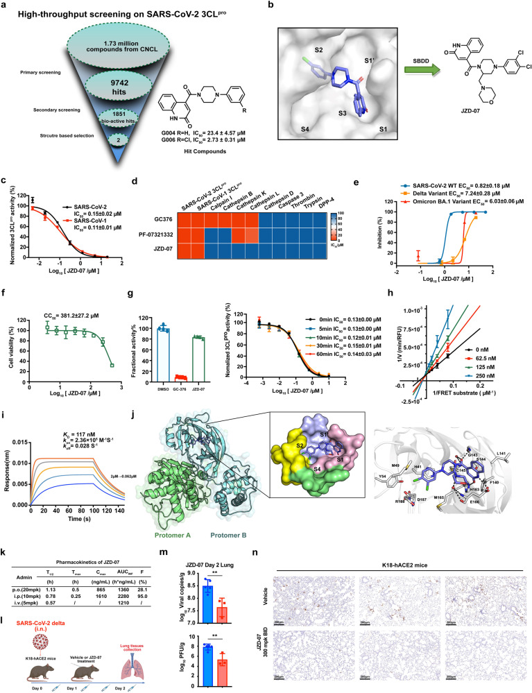
Fig. 1.
Discovery of Novel Non-peptidic and Non-covalent Small Molecule SARS-CoV‑2 3CL Protease Inhibitors. a Flowchart of the High-throughput screening and hit validation process. b Docking of lead compounds with SARS-CoV-2 3CLpro and rationale design for the noncovalent SARS-CoV-2 3CLpro inhibitor. c Compound JZD-07 is a potent inhibitor of SARS-CoV-2 3CLpro as well as SARS-CoV-1 3CL.pro d Biochemical assay of several human proteases, compound JZD-07 showing great selectivity against human protease, especially Calpain I and Cathepsin family protein, the values plotted were the IC50 values from the FRET-based enzymatic assay shown as a heatmap. e Inhibition of original strain (blue), Delta variant (yellow), and Omicron BA.1. variant (red) replication by Compound JZD-07 in Vero E6 cells. Data are shown as mean ± SD, n = 3 biological replicates. f Cytotoxicity (CC50) to Vero E6 cells measured by CCK-8 assays. Data are shown as mean ± SD, n = 3 biological replicates. g Rapid dilution assay and time-dependent inhibition of SARS-CoV-2 3CLpro by Compound JZD-07. A 40 nM SARS-CoV-2 3CLpro was preincubated with Compound JZD-07 for various periods of time (0 min to 1 h) before the addition of 20 μM FRET substrate to initiate the enzymatic reaction. Data are shown as mean ± SD. h Lineweaver-Burk plot depicting the mode of inhibition for Compound JZD-07 on SARS-CoV-2 3CLpro activity using different inhibitor concentrations. i Compound JZD-07 to SARS-CoV-2 3CLpro measured by biolayer interferometry (BLI), BLI analysis showing the representative association and disassociation curves of Compound JZD-07 at series concentrations. j Binding mode of JZD-07 (PDB ID: 8GTV) with SARS-CoV-2 3CLpro revealed by co-crystal structure. The two protomers are shown in the green (protomer A) and cyan (protomer B) cartoon, respectively. JZD-07 is shown as slate sticks. The surface representation of JZD-07 interacting with S1ʹ-S4 subsites of SARS-CoV-2 3CLpro. The subsites are colored light blue (S1ʹ), light pink (S1), yellow (S2), and green (S4). Interactions of JZD-07 with the surrounding residues are also revealed by the crystal structures. Residues (gray), as well as JZD-07, are shown in the sticks. H-bonds are represented by black dashed lines. k Preliminary PK evaluation of compound JZD-07 in mice. Dosed orally at 20 mg/kg. Dosed intraperitoneally at 10 mg/kg. Dosed intravenously at 5 mg/kg. l In vivo efficacy of compound JZD-07 in K18-hACE2 mice infected with SARS-CoV-2 delta variant. Experiment flow diagram. m Viral RNA copies and viral titers in lung tissues of each group were determined on day 2 after the SARS-CoV-2 delta variant challenge (**P ≤ 0.01). n Immunohistochemistry staining targets viral nucleocapsid protein. (SARS-CoV-2 Nucleocapsid Protein (HL344) Rabbit mAb #26369, CST). Viral nucleocapsid proteins were stained in brown

