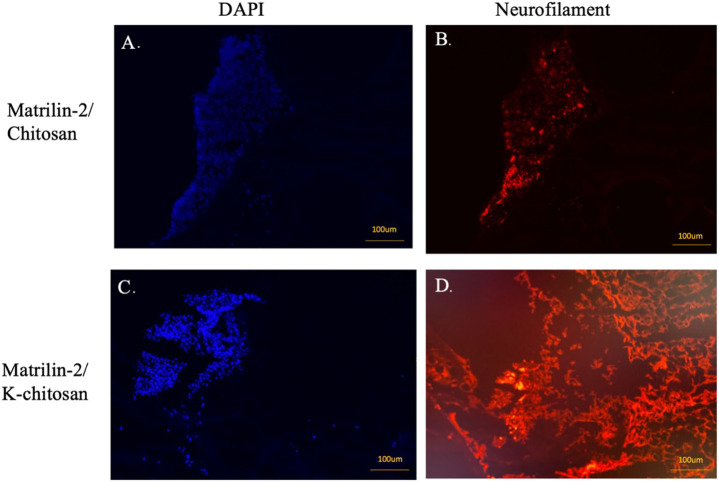
FIGURE 7.
Immunofluorescent imaging of matrilin-2/chitosan and matrilin-2/K-chitosan scaffold. (A) DAPI staining of neuronal cells adhered to end of conduit with matrilin-2/chitosan scaffold and (B) neurofilament staining of matrilin-2/chitosan demonstrating minimal axonal outgrowth from neuronal cells seen of same DAPI field. (C) DAPI staining of cluster of neuronal cells adhered to the end of conduit and matrilin-2/K-chitosan scaffold and (D) neurofilament staining of same DAPI field demonstrating axonal outgrowth. Scale marked at 100uM.

