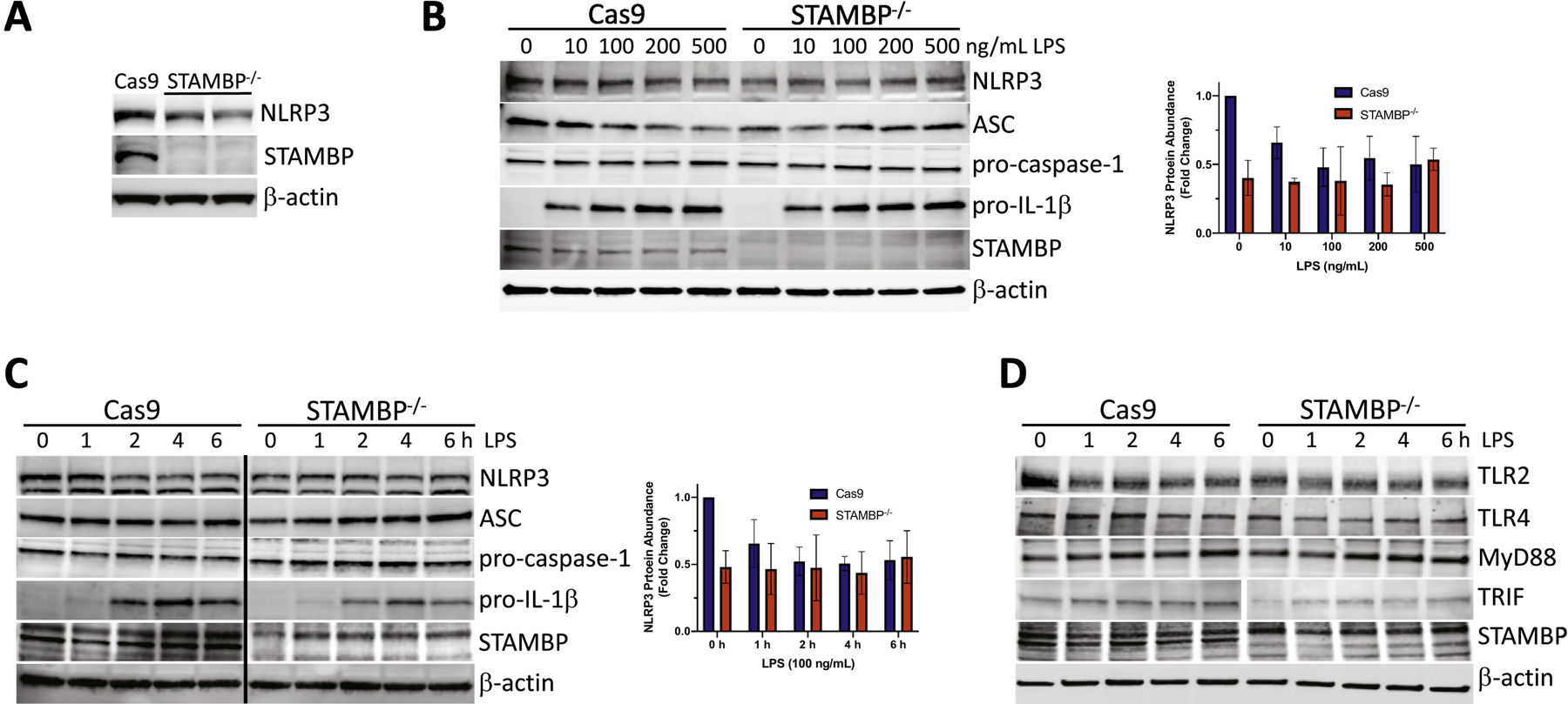
Figure 5: STAMBP knockout does not significantly impact cellular abundance of inflammasome proteins with endotoxin exposure.

(A) Baseline levels of NLRP3 were assayed by immunoblotting in STAMBP-/- THP-1 cells compared to Cas9 controls and demonstrate decreased basal NLRP3 protein abundance. (B-D) Cas9 control and STAMBP-/- THP-1 monocytes (1 x 10⁶ cells/mL) were exposed to LPS followed by immunoblotting. Inflammasome components NLRP3, ASC, pro-caspase-1 and pro-IL-1β were not significantly different in STAMBP-/- THP-1 monocytes compared to Cas9 THP-1 controls following exposure to (B) various concentrations of LPS for 16 hours or (C) following LPS exposure (100ng/mL) for the indicated time points. Graphs shown to right of immunoblots represent results of densitometic analysis of the NLRP3 signal, normalized to β-actin. Graphs represent mean ± SEM of three separate experiments. (D) Immunoreactive levels of upstream NLRP3 activating proteins TLR2, TLR4, MyD88 and TRIF were not significantly different in STAMBP-/- THP-1 cells compared to Cas9 controls following exposure to LPS (100ng/mL) for the indicated time points.
