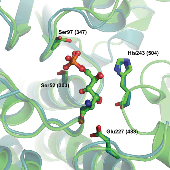
FIGURE 4.

Superposition of the structures of the active sites of 1MOS (E. coli GFAT) (deep teal) and 8EOL (S. denitrificans NagB‐II) (green), showing a molecule of GlcNol6P bound to the active site pocket in both enzymes. Residues His243 (504), Ser52 (303), Ser97 (347), and Glu227 (488) are shown in stick representation, and GlcNol6P molecules are in ball and stick representation. The numbers in parentheses are for GFAT.
