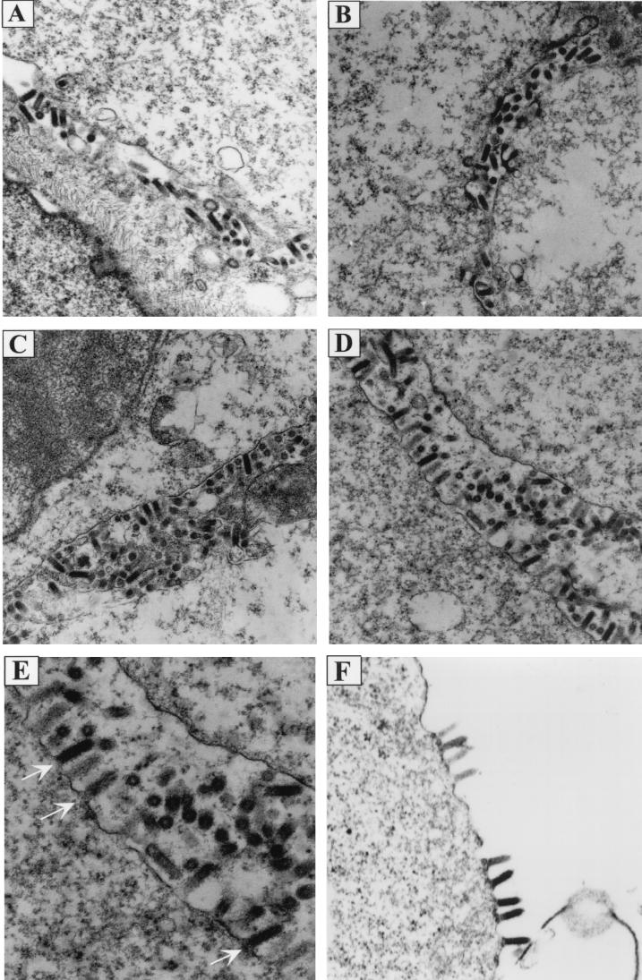
FIG. 5.
Electron micrographs showing morphologies of PPXY mutants. BHK-21 cells infected with WT or mutant viruses were fixed at 8 h p.i., and thin sections of cells were examined under the electron microscope to determine the stage(s) at which morphogenesis of PPXY mutants was blocked. (A) WT; (B) ΔG-GFP; (C) AAPY; (D and E) PPPA; (F) ΔG-AAPA. Magnifications: ×15,000 for panels A through D; ×18,000 for panel F. Panel E) is an enlargement of panel D. Arrows in panel E point to the budding virions attached to the plasma membrane of the infected cell.

