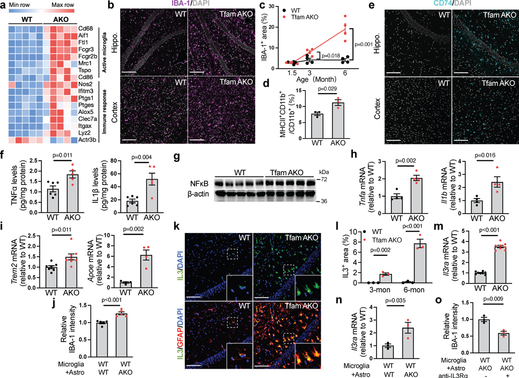
Fig. 6. Astrocytes with OxPhos deficit promote microglial activation and neuroinflammation via IL-3.
(a) Heatmap showing DEGs (FDR-corrected p < 0.05) related to microglial activation and immune response in 6-month mouse hippocampi. (b) Representative images of hippocampal or cortical sections of 6-month mice stained for IBA-1. (c) IBA-1+ area % in hippocampal sections of 1.5-, 3-, and 6-month WT and TfamAKO mice. (d) MHCII-positive rate in microglia acutely isolated from 6-month mouse brains with anti-CD11b microbeads. (e) Representative images of hippocampal or cortical sections of 6-month mice stained for CD74. (f) TNFα and IL-1β levels in the cortex of 6-month mice. (g) Western blots of NFκB in 6-month mouse hippocampi (quantified in Extended Data Fig. 7e). (h and i) mRNA level of Tnfa, Il1b, Trem2, and Apoe in microglia acutely isolated from 6-month mouse brains. (j) IBA-1 intensity (normalized to cell count) in WT primary microglia (on coverslips in 6-well plates) cocultured with WT or TfamAKO astrocytes (in 6-well inserts) for 24 h. (k) Representative images of hippocampal sections of 6-month mice stained for IL-3 and GFAP. (l) IL-3+ area in the hippocampus of 3- and 6-month mice. (m) Il3ra mRNA levels in microglia acutely isolated from 6-month mouse brains. (n) Il3ra mRNA levels in WT microglia (in 24-well plates) cultured with WT or TfamAKO astrocytes (in 24-well inserts) for 24 h. (o) IBA-1 intensity (normalized to cell count) in WT microglia (on coverslips in 6-well plates) pretreated with vehicle or IL-3Rα neutralizing antibody before being cocultured with TfamAKO astrocytes (in 6-well inserts) for 24 h. n = 4 (c, h), 5 (j), 3 (d, l, n, o), or 8 (m) mice or independent samples; n = 6 (WT) or 5 (AKO) mice (f); n = 7 (Trem2-WT), 8 (Trem2-AKO), or 4 (Apoe) mice (i). Bar graphs and dot plots are presented as mean ± SEM. Two-sided unpaired t-test was used for all comparisons. Scale bars, 100 μm (b, e, and k).

