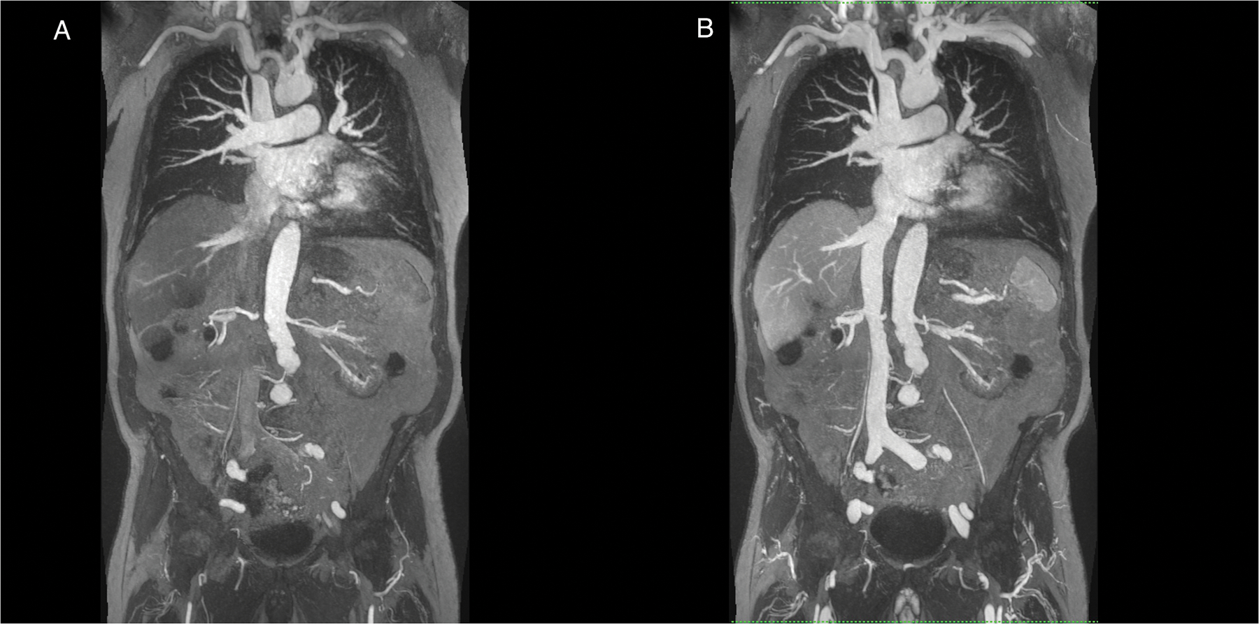
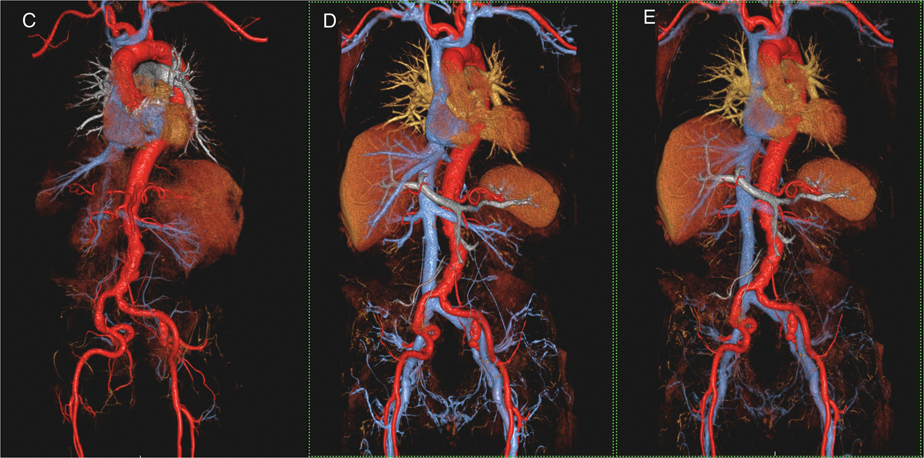
Figure1 (A-E).


A 90 year old male patient with renal failure undergoing vascular evaluation for transcatheter aortic valve replacement (TAVR). First pass imaging with ferumoxytol (A,C) on thin maximum intensity projection (MIP) (A) and volume rendered (VR) (C) reconstructions show similar bright and uniform arterial enhancement as on steady state images (B,D,E), where arteries and veins show equal signal intensity. In E, the systemic veins have been made more transparent. Note extensive irregularity and tortuosity in the aorto-iliac vessels. This study was acquired at 3.0T.
