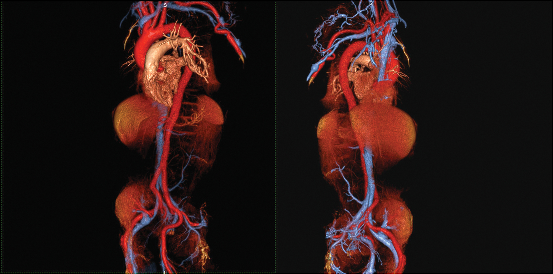
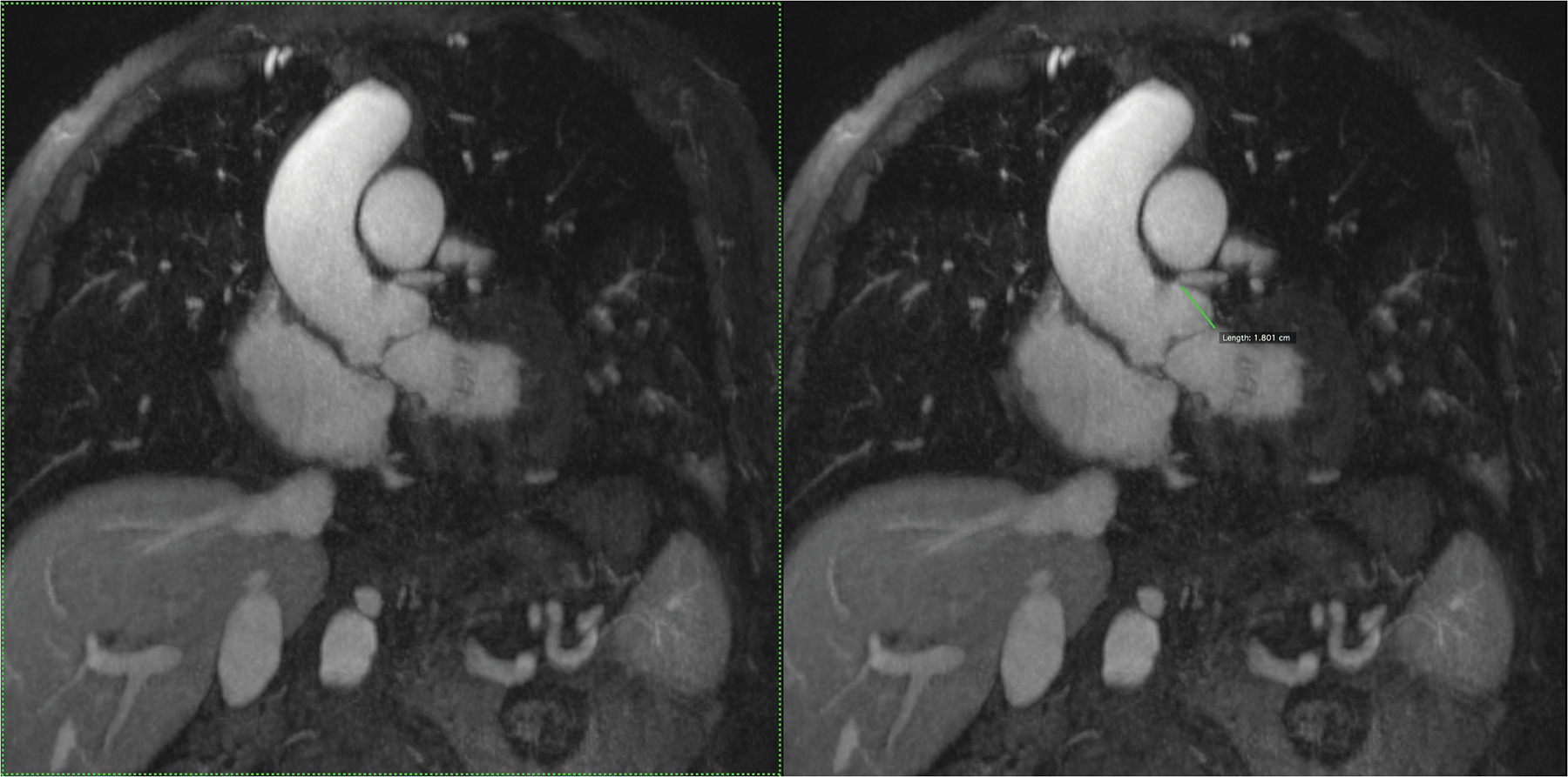
Figure 4 (A,B).


An 87 year old male with aortic stenosis, sinus bradycardia and renal transplant undergoing evaluation for TAVR placement. Volume rendered images in the steady state distribution of ferumoxytol (A) display the entire aorto-iliac anatomy as well as the patent renal transplant artery and vein in the pelvis. Gated, spoiled gradient echo cine images show the aortic valve leaflets and the relationship of the left coronary artery ostium to the annulus (B).
