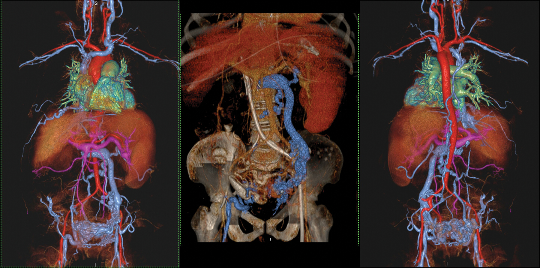
Figure 5.

A 46 year old female with end stage renal failure and liver disease with extensive venous thrombosis. Undergoing evaluation for organ transplantation. Volume rendered acquisition with steady state ferumoxytol (left and right frames) show detailed vascular anatomy of the thorax, abdomen and pelvis, including a hugely dilated left gonadal vein and pelvic varices. A CT venogram (middle frame) performed shortly prior to MRI suffers from contrast dilution with poorer vascular definition. Note the left sided venous catheter extending proximally through the occluded IVC. The MRI study was acquired at 3.0T.
