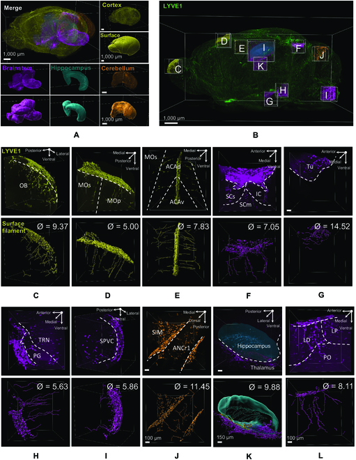
Fig. 2.

Characteristics of deep brain lymphatic vessels. (A) and (B) 3D reconstruction of whole brain with 3D reconstruction of the selected and analyzed brain regions. Different brain regions highlighted in (A) and (B) are color-coded throughout all the images below. (C to L) Top: Original LYVE1 signals. Bottom: IMARIS processed images by using Filament and Surface algorithm showing the origin of the deep brain lymphatic vessels in each brain region and their branching, direction, and diameter (Ø). OB, olfactory bulb; MOs, secondary motor cortex; MOp, primary motor cortex; ACAd, anterior cingulate cortex dorsalis; ACAv, anterior cingulate cortex ventralis; LD, lateral dorsal nucleus of thalamus; LP, lateral posterior nucleus of the thalamus; PO, posterior complex of the thalamus; Tu, tuberal nucleus; PG, pontine gray; SPVC, caudal part of spinal nucleus of the trigeminal; TRN, tegmental reticular nucleus (all in brainstem); SCs, sensory superior colliculus; SCm, motor superior colliculus; IC, inferior colliculus; SIM, simple lobule (in midbrain); ANCr1, ansiform lobule crus 1 (in cerebellum).
