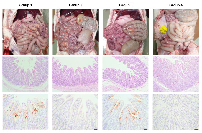
Fig. 4.
Macroscopic and microscopic small intestine lesions in piglets after inoculation with PEDV (group 1), CPA (group 2), or PEDV/CPA (group 3), and in sham control animals (group 4). Small intestines of individual piglets from each group were examined for gross lesions. Representative necropsy images are presented at the top of each panel. Hematoxylin- and eosin-stained tissue sections of the proximal jejunum from representative piglets in each group are shown at the middle of each panel (100× magnification, scale bar = 100 µm). IHC analysis results showing PEDV antigens in jejunal tissue sections from representative pigs in each group are presented at the bottom of each panel (200× magnification, scale bar = 50 µm). Immunostaining of the PEDV N proteins (brown staining) was detected in the epithelial cells of the proximal jejunum in piglets inoculated with PEDV alone and in those co-inoculated with CPA and PEDV. No PEDV antigen was identified in the small intestine of the CPA single-inoculated and mock-inoculated piglets.

