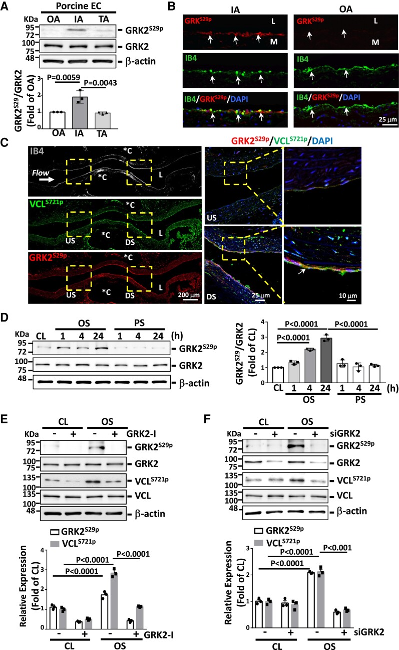
Figure 2.
Disturbed flow induction of VCLS721p is mediated by GRK2S29p. (A) Western blot analysis of the expression of indicated proteins in fresh porcine aortic ECs. (B) Immunofluorescence staining for GRK2S29p expression in the porcine IA and OA EC layers. Arrows indicate the EC layer stained for IB4. (C) Longitudinally panoramic examination of EC GRK2S29p and VCLS721p expressions from the upstream (US) through the midpoint to the downstream (DS) areas of the constriction in the experimentally stenosed rat abdominal aorta. Co-immunostaining for GRK2S29p and VCLS721p indicates their co-localization in ECs (arrows). *C indicates clip-injured areas. Right panels show the magnified views of the indicated areas (dashed line box). (D–F) ECs were kept under static condition as controls (CL) or subjected to oscillatory (OS) or pulsatile (PS) shear stress for the indicated times (D) or 24 h (E and F). In some experiments, ECs were pre-treated with GRK2-specific inhibitor (GRK2-I, 30 μM) (E) or transfected with GRK2-specific siRNA (siGRK2, 15 nM) (F) for 24 h. Data in A, D–F are means ± SEM (n = 3). One-way ANOVA with the Tukey multiple comparison test was applied to A and D. Two-way ANOVA with the Tukey multiple comparison test was applied to E and F. Results in each figure represent three independent experiments with similar results.

