Figure 3.
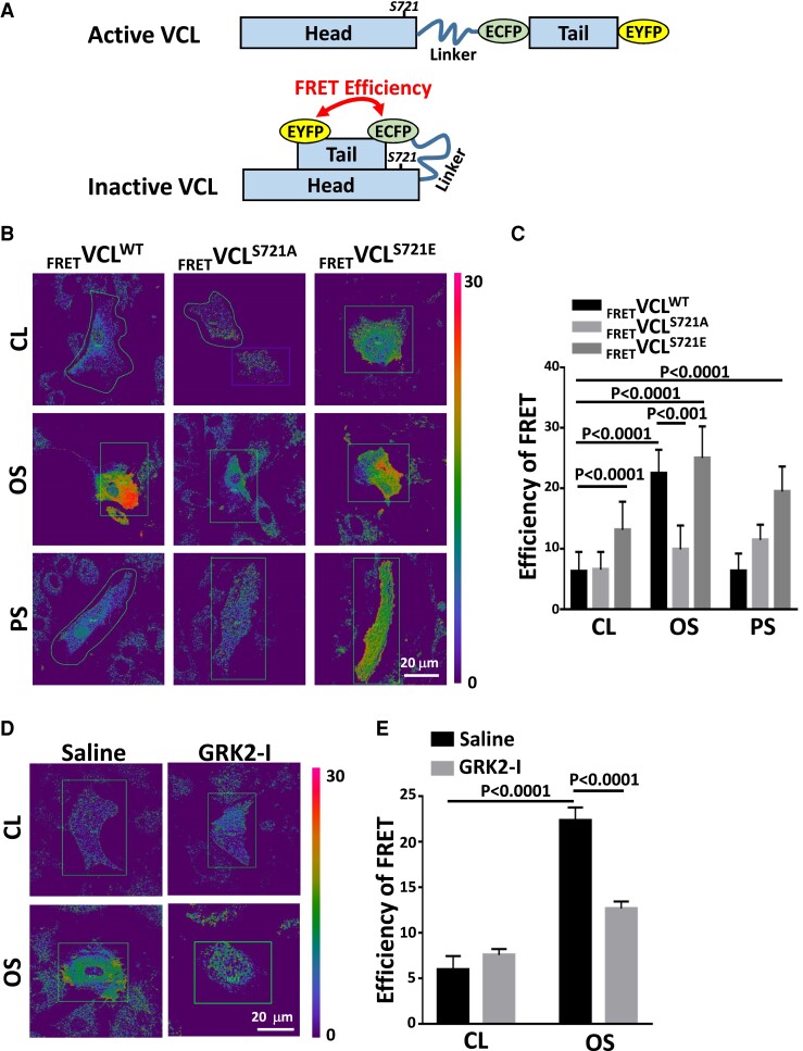
Disturbed flow induces an inactive form of VCL with a closed conformation. (A) Schematic diagrams showing the conformational changes of the VCL conformation-responsive FRET construct used in this study. Active VCL exhibits an open conformation in which intramolecular head–tail interaction is severed. Inactive VCL shows a closed conformation characterized by a tight head–tail interaction. Inactive VCL induces a high FRET signal, whereas the FRET signal is diminished for active VCL. (B and C) ECs were transfected with wild-type VCL FRET probe (FRETVCLWT) and mutant VCL FRET probes (FRETVCLS721A and FRETVCLS721E) for 24 h and then kept under static condition as controls (CL) or subjected to oscillatory (OS) or pulsatile (PS) shear stress for 24 h. Pseudocolor FRET ratio images of these VCL FRET probes in live ECs were corrected after acceptor photobleaching (B), and the extracted FRET efficiencies were quantified (n = 25) (C). (D and E) FRETVCLWT-transfected ECs were incubated with GRK2 inhibitor (GRK2-I, 30 μM) or control saline for 24 h. These cells were kept under static condition as controls (CL) or subjected to oscillatory shear stress (OS) for 24 h, and their FRET pseudocolor signals were examined. The mean values of extracted FRET efficiency were quantified (n = 15) (E). Data in C and E are means ± SEM from three independent experiments and were analyzed by two-way ANOVA with Tukey multiple comparison test. The images in the figure represent three independent experiments with similar results.

