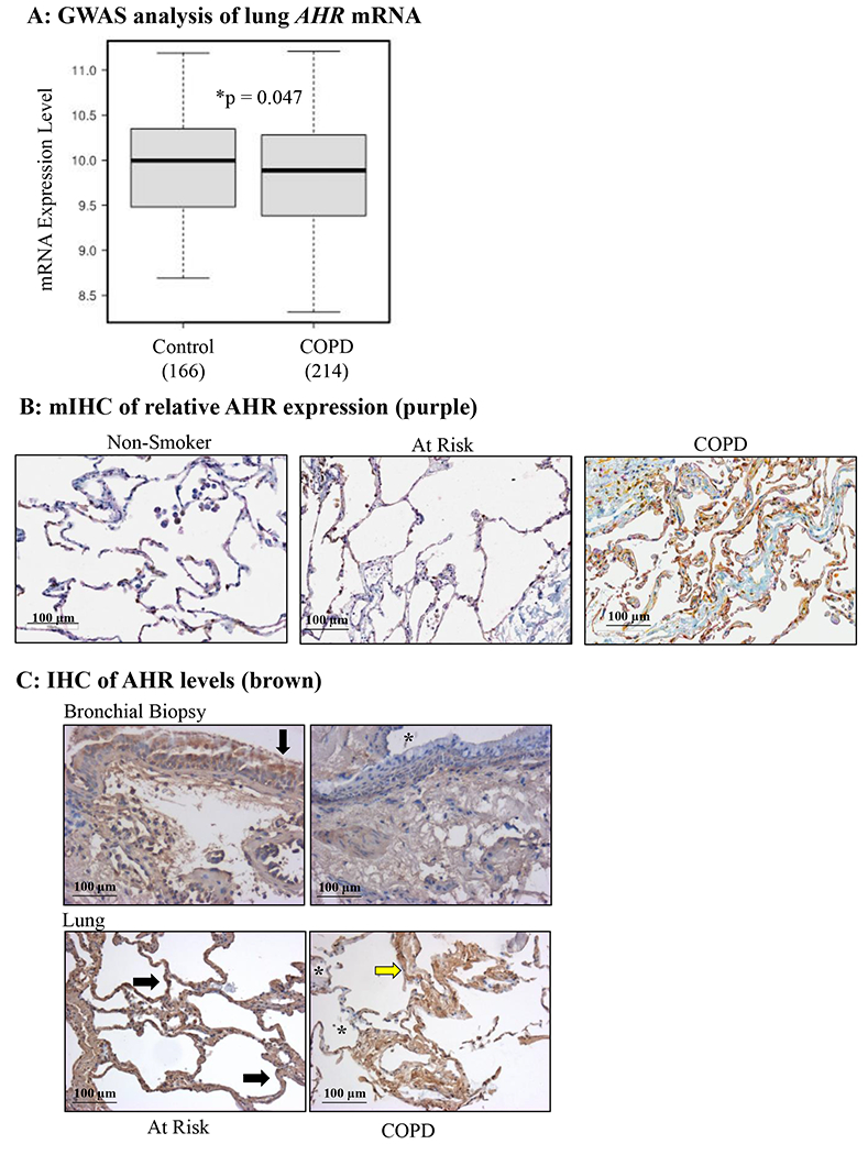
FIGURE 3.

AHR expression is reduced in lung tissue from human COPD subjects. A: There was significantly less AHR mRNA in the lungs of COPD subjects (n=214) compared to subjects without COPD (Control) (n=166). B: mIHC was used to detect AHR (purple), vimentin (fibroblasts; yellow) and cytokeratin-19 (epithelial cells; brown) in human lungs. Note the relative decrease in AHR (purple) in COPD subjects relative to Non-smokers and those At Risk (smokers without COPD); representative images are shown. C: Bronchial biopsy (top panels) and lung tissue (bottom panels)- there is less intense AHR expression (brown) in lungs tissue from COPD subjects compared to those considered At Risk (smokers); regions of noticeably less AHR are denoted by an asterisk (*). Representative images are shown.
