Figure 3.
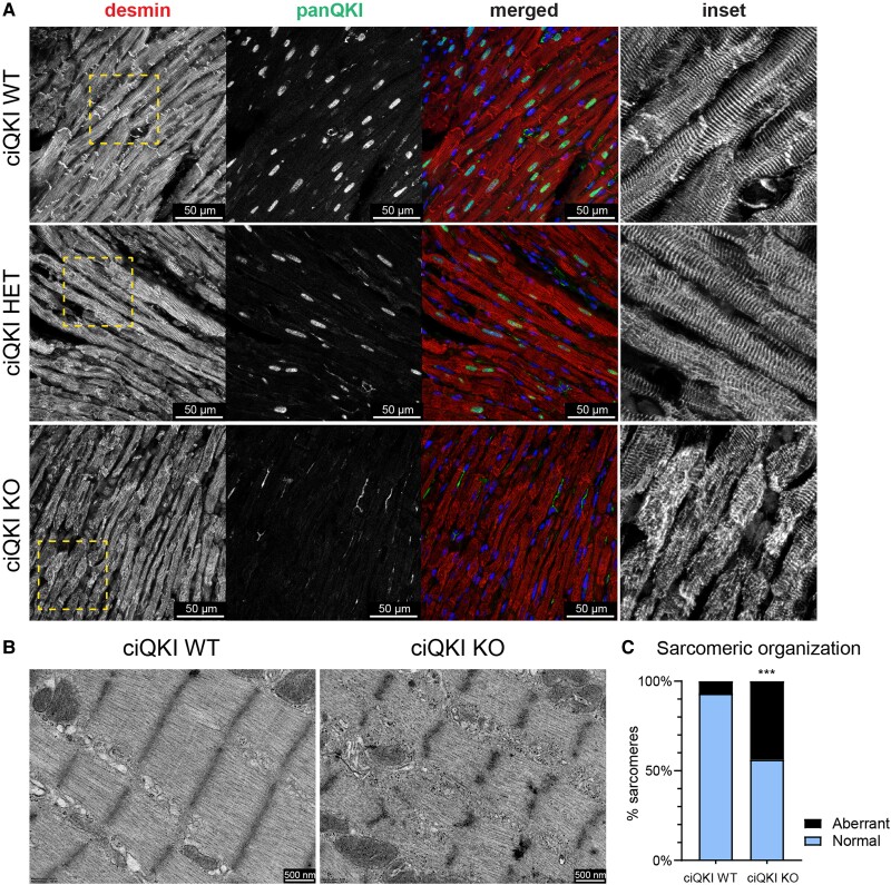
Loss of QKI disrupts cytoskeletal and sarcomere organization in the adult heart. (A) Immunocytochemistry showing the cytoskeletal protein desmin (red), panQKI (green), and DAPI (blue). The right panels (inset) represent magnifications of the yellow box in the desmin panels. Scale bar: 50 µm. (B) Representative transmission electron microscopy images of cardiomyocytes from ciQKI WT and ciQKI KO mice. Scale bar: 500 nm. (C) Quantification of aberrant sarcomeres in LV cardiomyocytes in electron microscopy images. Three mice per group and four images per mouse. χ2 test. ***adjusted P < 0.001.

